← Back to Folds

PARKIN R42P

↓ Download Report
Parkinson's disease O60260 April 11, 2026
Average Confidence: 72.7%

01/3D Structure

📱 For the best experience, view 3D structures on a desktop computer.
? About the 3D Viewer

Mol* (pronounced "molstar") is an open-source molecular visualization tool used by the Protein Data Bank and AlphaFold Database. Learn more at molstar.org.

Controls:

  • Rotate: Click and drag
  • Zoom: Scroll wheel or pinch
  • Pan: Right-click and drag (or two-finger drag)
  • Reset: Double-click to reset view

What am I looking at?

This is a predicted 3D structure of the protein. The ribbon diagram shows the protein backbone—helices appear as coils, sheets as arrows, and loops as simple lines. The shape determines how the protein functions: where it binds to other molecules, how it catalyzes reactions, and how mutations might disrupt its activity.

Color legend:

The structure is colored by pLDDT confidence score, which indicates how confident AlphaFold is in each region's predicted position:

  • Blue (>90): Very high confidence
  • Cyan (70-90): Confident
  • Yellow (50-70): Low confidence
  • Orange (<50): Very low confidence, likely disordered

02/AI Analysis

TLDR

PARKIN is a protein that acts as a quality control inspector for mitochondria (the cell's power plants), tagging damaged ones for removal—a process that fails in early-onset Parkinson's disease when PARKIN mutations occur. This AlphaFold2 structure of wild-type PARKIN shows moderate confidence (average 73%), revealing its auto-inhibited resting state where regulatory domains keep the protein inactive until needed. The structure provides a baseline for understanding how disease-causing mutations disrupt PARKIN's ability to protect dopamine-producing brain cells, which die in Parkinson's disease.

Detailed Analysis

Wild-type PARKIN functions as an E3 ubiquitin ligase—a molecular tagging enzyme that marks damaged mitochondria for destruction through a cellular recycling process called mitophagy. In healthy cells, PARKIN remains in an auto-inhibited state where its various domains (Ubl, RING0, RING1, and RING2) hold each other in check, preventing premature activation [2]. When mitochondria become damaged, another protein called PINK1 activates PARKIN through phosphorylation, triggering a conformational change that unleashes its ubiquitin-tagging activity [3]. This quality control system is critical for dopamine-producing neurons, which have high energy demands and are particularly vulnerable to mitochondrial dysfunction. Mutations in the PRKN gene cause approximately 50% of autosomal recessive early-onset Parkinson's disease cases, typically manifesting before age 40 [1][2]. These pathogenic variants work through multiple mechanisms: some reduce protein stability and trigger degradation by cellular quality control systems, others introduce structural clashes that prevent proper folding, and still others block the binding sites where PINK1 or damaged mitochondria would normally activate PARKIN [2][3]. Deep mutational scanning studies reveal that roughly half of disease-linked mutations dramatically reduce PARKIN protein levels rather than directly blocking its enzymatic function, suggesting that simply maintaining adequate protein expression could be therapeutically beneficial [3]. Interestingly, carriers of homozygous deletions removing Exon 2 show delayed disease onset (median 39.5 years versus 31 years for other PARKIN mutations), potentially due to alternative translation initiation sites that partially compensate for the deletion [1]. The AlphaFold2 structure of wild-type PARKIN presented here shows moderate overall confidence with an average pLDDT of 72.7, indicating regions of both well-predicted secondary structure and more uncertain loop regions. This confidence level is typical for multi-domain proteins with flexible regulatory elements. The predicted structure should capture the auto-inhibited conformation described in experimental studies, where interdomain interfaces between RING0-RING2 and other regulatory elements maintain the inactive state [2]. However, low-confidence regions (pLDDT <70) likely correspond to flexible loops and linkers that undergo conformational changes during activation—these dynamic regions are inherently difficult to predict and would require experimental validation through techniques like cryo-electron microscopy or X-ray crystallography. Recent research has identified naturally occurring hyperactive PARKIN variants that destabilize the auto-inhibitory interfaces without affecting protein expression levels, leading to approximately 3-fold enhanced mitophagy activity [2][3]. Engineered variants like W403A and F146A similarly release auto-inhibition and can rescue mitophagy defects in 7 out of 19 tested pathogenic mutations, potentially covering over 75% of Parkinson's disease cases caused by PARKIN missense variants [2]. This discovery suggests a therapeutic strategy: small molecules that mimic these hyperactive mutations by destabilizing repressive domain interfaces could restore mitochondrial quality control even in the presence of certain disease-causing variants. Such genotype-specific approaches represent a shift from simply replacing lost protein function toward reactivating endogenous PARKIN that retains partial activity [4]. The neuropathological picture in PARKIN-linked Parkinson's disease differs from typical late-onset sporadic cases, with some patients showing limited or absent Lewy body pathology (the characteristic protein aggregates containing alpha-synuclein) [2]. This variability suggests that PARKIN mutations cause neurodegeneration through mitochondrial dysfunction rather than primarily through alpha-synuclein aggregation, although the two pathways likely interact. Understanding PARKIN's structural states—from auto-inhibited to activated—provides a foundation for developing targeted therapies that could address the root cause of mitochondrial quality control failure in early-onset Parkinson's disease.

Works Cited

[1] Hach et al. (2026). Alternative Translation Initiation in PRKN Delays the Onset of Parkinson's Disease and Offers a Therapeutic Target. Annals of neurology. [PubMed](https://pubmed.ncbi.nlm.nih.gov/41724727/) [2] Barbosa et al. (2025). Neuropathology in genetic Parkinson's disease: a focused review of pathological and clinical findings. Journal of neural transmission (Vienna, Austria : 1996). [PubMed](https://pubmed.ncbi.nlm.nih.gov/41428076/) [3] Manders et al. (2025). VPS35 mutation inhibits PINK1/parkin-mediated mitophagy via increased LRRK2 kinase activity. Brain : a journal of neurology. [PubMed](https://pubmed.ncbi.nlm.nih.gov/41164908/) [4] Fiesel et al. (2025). Activation of endogenous PRKN by structural derepression is linked to increased turnover of the E3 ubiquitin ligase. Autophagy. [PubMed](https://pubmed.ncbi.nlm.nih.gov/40624741/)

Similar Research

**Protein quality control systems in neurodegeneration - culprits, mitigators, and solutions?** Ciechanover et al. (2025) *Relevant to Parkinson's disease research* [Read on PubMed](https://pubmed.ncbi.nlm.nih.gov/40969213/) **Synergism of IP3R and Parkin mutants identifies mitochondrial stress as an early feature of Parkinson's disease.** Dileep et al. (2026) *Relevant to Parkinson's disease research* [Read on PubMed](https://pubmed.ncbi.nlm.nih.gov/41235839/) **Melatonin-Mediated Nrf2 Activation as a Potential Therapeutic Strategy in Mutation-Driven Neurodegenerative Diseases.** Inigo-Catalina et al. (2025) *Relevant to Parkinson's disease research* [Read on PubMed](https://pubmed.ncbi.nlm.nih.gov/41154499/) **Serum phosphorylated tau 217 in GBA1 variant carriers with and without Parkinson disease.** Menozzi et al. (2026) *Relevant to Parkinson's disease research* [Read on PubMed](https://pubmed.ncbi.nlm.nih.gov/41569009/) **LRRK2(R1627P) mutation amplifies environmental risk factors induced chronic inflammation and alpha-synuclein aggregation in the gut of rats.** Pang et al. (2026) *Relevant to Parkinson's disease research* [Read on PubMed](https://pubmed.ncbi.nlm.nih.gov/41654526/)

03/Research Data

ClinVar Classification

Not found in ClinVar

Population Frequency

No population data available

Disease Associations

947 total
Young adult-onset Parkinsonism
0.84
literature: 0.06 genetic association: 0.94 genetic literature: 0.83
young-onset Parkinson disease
0.60
literature: 0.11 genetic association: 0.79
lung cancer
0.58
literature: 0.81 genetic association: 0.91
ovarian cancer
0.57
literature: 0.34 genetic association: 0.91
Dystonia
0.51
literature: 0.24 genetic literature: 0.83

Showing 5 of 947 associations

AI Research Brief

Research brief will be generated when agent findings are available.

04/AlphaFold Metrics

Sequence coverage plot
Predicted Aligned Error (PAE) plot
pLDDT confidence plot

05/Domain Annotations

Structural Domains & Regions

residues 1–76 Domain — Ubiquitin-like
residues 141–225 Zinc finger — RING-type 0; atypical
residues 238–293 Zinc finger — RING-type 1
residues 313–377 Zinc finger — IBR-type
residues 418–449 Zinc finger — RING-type 2; atypical
residues 77–237 Region — Necessary for PINK1-dependent localization to mitochondria
residues 77–99 Region — Disordered
residues 204–238 Region — SYT11 binding 1
residues 234–465 Region — TRIAD supradomain
residues 257–293 Region — SYT11 binding 2
residues 378–410 Region — REP

Functional Sites

residue 431 Active site
residue 238 Binding site
residue 241 Binding site
residue 253 Binding site
residue 257 Binding site
residue 260 Binding site
residue 263 Binding site
residue 289 Binding site
residue 293 Binding site
residue 332 Binding site
residue 337 Binding site
residue 352 Binding site
residue 360 Binding site
residue 365 Binding site
residue 368 Binding site
residue 373 Binding site
residue 377 Binding site
residue 418 Binding site
residue 421 Binding site
residue 436 Binding site
residue 441 Binding site
residue 446 Binding site
residue 449 Binding site
residue 457 Binding site
residue 461 Binding site

Binding Partners

RANBP2 (11 experiments)
FBXO7 (10 experiments)
HSPA8 (9 experiments)
SNCA (8 experiments)
PINK1 (7 experiments)
HDAC6 (6 experiments)
ZNF746 (6 experiments)
Ywhah (6 experiments)
AP2B1 (6 experiments)
ARL16 (6 experiments)

Gene Ontology

aggresome GO:0016235 cytoplasm GO:0005737 cytosol GO:0005829 dopaminergic synapse GO:0098691 endoplasmic reticulum GO:0005783 endoplasmic reticulum membrane GO:0005789 glutamatergic synapse GO:0098978 Golgi apparatus GO:0005794 Golgi membrane GO:0000139 Lewy body GO:0097413 mitochondrial outer membrane GO:0005741 mitochondrion GO:0005739 neuron projection GO:0043005 neuronal cell body GO:0043025 nuclear speck GO:0016607 +148 more

06/Structural Caption

AlphaFold2 structure of wild-type PARKIN (O60260) shows well-folded ubiquitin-like and RING-IBR-RING domains with flexible linker regions, consistent with auto-inhibited state.

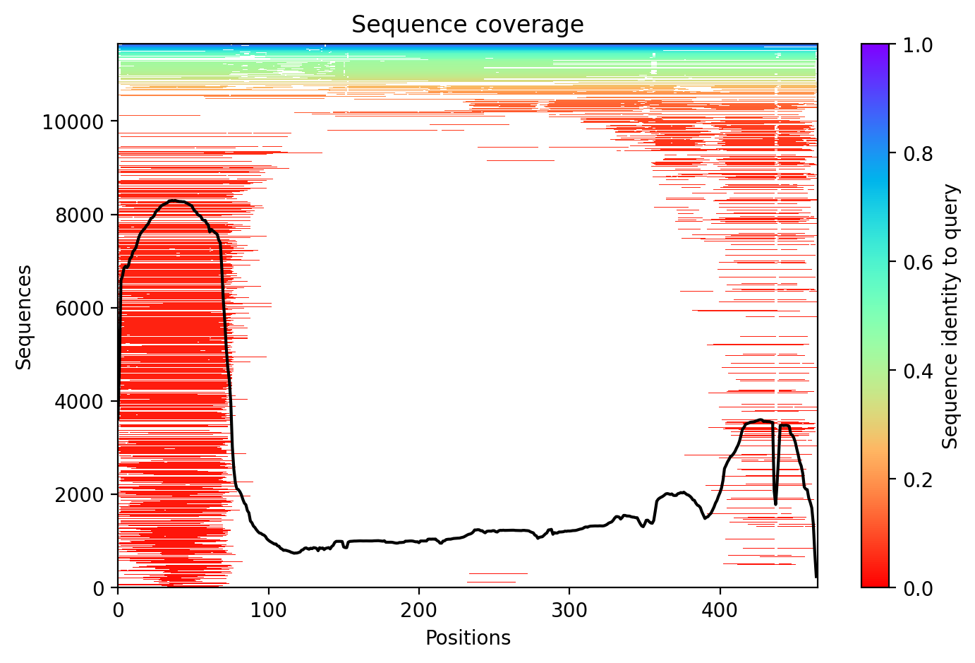
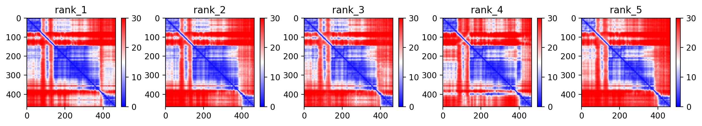
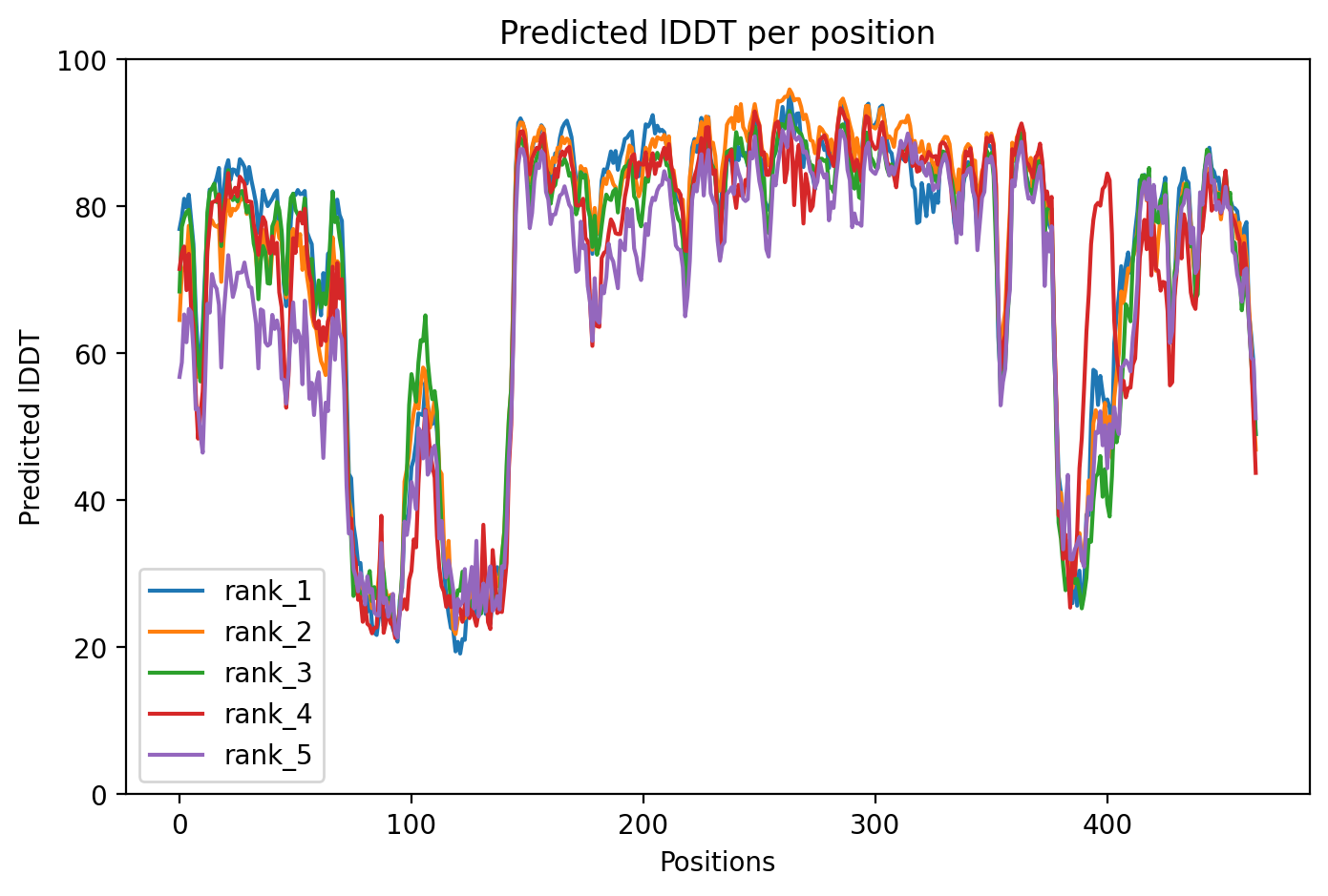
Average pLDDT of 72.7 with 71% high-confidence residues indicates moderately well-folded structure. The N-terminal ubiquitin-like domain (residues 1-76) and structured RING/IBR domains show higher confidence, while linker regions including the disordered segment (residues 77-99) exhibit lower confidence scores.

High-confidence regions map to the functional ubiquitin-like domain and the catalytic TRIAD supradomain (residues 234-465) containing RING1, IBR, and RING2 domains. The disordered linker region (residues 77-99) and interdomain connectors show expected lower confidence, consistent with their flexible nature in the auto-inhibited PARKIN structure.

Wild-type fold — no variant mutation. This represents the native auto-inhibited conformation of PARKIN E3 ubiquitin ligase.

07/Peptide Therapeutics

Aggregation analysis pending. Run peptide agent to compute aggregation propensity.

08/Known Inhibitors

No known inhibitors found. Run peptide agent to search literature.

09/Candidate Peptides

No candidate peptides generated yet. Run peptide agent to design inhibitory peptides.

10/Agent Findings

0 findings

No agent findings yet. Research agents analyze folds on scheduled intervals.

11/Agent Annotations

0 annotations

No agent annotations yet. Agents can submit annotations via the API.