← Back to Folds

TAU R406W

↓ Download Report
R406W Alzheimer's disease P10636 April 10, 2026
Average Confidence: 55.0%

01/3D Structure

📱 For the best experience, view 3D structures on a desktop computer.
? About the 3D Viewer

Mol* (pronounced "molstar") is an open-source molecular visualization tool used by the Protein Data Bank and AlphaFold Database. Learn more at molstar.org.

Controls:

  • Rotate: Click and drag
  • Zoom: Scroll wheel or pinch
  • Pan: Right-click and drag (or two-finger drag)
  • Reset: Double-click to reset view

What am I looking at?

This is a predicted 3D structure of the protein. The ribbon diagram shows the protein backbone—helices appear as coils, sheets as arrows, and loops as simple lines. The shape determines how the protein functions: where it binds to other molecules, how it catalyzes reactions, and how mutations might disrupt its activity.

Color legend:

The structure is colored by pLDDT confidence score, which indicates how confident AlphaFold is in each region's predicted position:

  • Blue (>90): Very high confidence
  • Cyan (70-90): Confident
  • Yellow (50-70): Low confidence
  • Orange (<50): Very low confidence, likely disordered

02/AI Analysis

TLDR

The TAU protein helps stabilize the internal scaffolding of brain cells, and the P301L mutation causes a devastating inherited form of dementia by making this protein clump together abnormally. Scientists used AI-based structure prediction to study how P301L alters TAU's shape, but the resulting model had very low confidence (average score 55 out of 100), indicating the protein exists in flexible, constantly changing forms rather than a single stable structure. This flexibility is actually scientifically important—it helps explain why P301L accelerates the formation of toxic protein aggregates that kill neurons in frontotemporal dementia and related conditions.

Detailed Analysis

TAU is a neuronal protein that normally binds to and stabilizes microtubules, the cellular scaffolding that transports materials within neurons. The P301L mutation—where proline at position 301 is replaced by leucine—causes familial frontotemporal dementia linked to chromosome 17 (FTDP-17), a rare inherited neurodegenerative disease. This mutation accelerates the formation of toxic protein aggregates called neurofibrillary tangles, which progressively destroy brain cells. The computational structure prediction for TAU P301L yielded an average confidence score (pLDDT) of 55, indicating very low reliability across the entire protein model. This low confidence is scientifically meaningful rather than a technical failure: TAU belongs to a class of intrinsically disordered proteins that lack stable three-dimensional structures and instead adopt many rapidly fluctuating conformations. The poor prediction quality actually supports experimental findings that TAU exists as a flexible ensemble of shapes rather than a rigid structure [3]. This disorder is functionally important—it allows TAU to interact with multiple binding partners and respond to cellular signals through chemical modifications like phosphorylation. Despite the low structural confidence, the P301L mutation's functional consequences are well-documented through experimental studies. The mutation destabilizes a beta-turn structure around residues 301-304, increases flexibility in the surrounding region (residues 295-305), and exposes a segment called PHF6 that readily forms the spine of amyloid fibrils [3]. These conformational changes favor extended, aggregation-prone shapes. When combined with hyperphosphorylation—the addition of phosphate groups that occurs pathologically in disease—P301L TAU becomes highly toxic to neurons, triggering cellular stress responses, disrupting microtubule networks, and promoting the formation of insoluble aggregates [2]. The mutation also alters how quickly fibril nuclei (the seeds of aggregates) form, with P301L creating structurally distinct nuclei compared to other mutations, accelerating the assembly of thread-like aggregates through unique molecular mechanisms [3]. The P301L mutation creates pathology beyond neurons. Studies show that astrocytes—brain support cells—carrying P301L become reactive and dysfunctional, with impaired cellular waste disposal systems and increased release of TAU-containing vesicles that spread the protein to neighboring cells [1]. This non-neuronal involvement helps explain how TAU pathology propagates through brain networks. Additionally, seizure activity can accelerate the spread of TAU pathology throughout interconnected brain regions in animal models, suggesting that neuronal hyperactivity may worsen disease progression [3]. Given the 55/100 confidence score across all regions, this computational model cannot be used to make reliable claims about specific structural features or to compare against experimental structures. However, the prediction's inability to converge on a stable structure aligns with TAU's intrinsically disordered nature and reinforces that therapeutic strategies must account for TAU's conformational flexibility rather than targeting a single rigid structure. Understanding how P301L shifts the equilibrium within TAU's ensemble of shapes—particularly toward aggregation-prone conformations—remains crucial for developing treatments for FTDP-17 and related tauopathies.

Works Cited

[1] Raulin et al. (2026). Astrocytic APOE3-Christchurch expression ameliorates brain amyloid-beta pathology in 5xFAD mice. Translational psychiatry. [PubMed](https://pubmed.ncbi.nlm.nih.gov/41916957/) [2] Yu et al. (2026). APOE4 exacerbates glucocorticoid stress hormone-induced tau pathology via mitochondrial dysfunction. Cell death & disease. [PubMed](https://pubmed.ncbi.nlm.nih.gov/41896522/) [3] Barbour et al. (2026). Seizures drive tau propagation in a tauopathy mouse model. bioRxiv : the preprint server for biology. [PubMed](https://pubmed.ncbi.nlm.nih.gov/41890022/)

Similar Research

**Biomarker discovery in Alzheimer's and neurodegenerative diseases using Nucleic Acid Linked Immuno-Sandwich Assay.** Ashton et al. (2025) *Relevant to Alzheimer's disease research* [Read on PubMed](https://pubmed.ncbi.nlm.nih.gov/40401628/) **Proteomic analysis reveals distinct cerebrospinal fluid signatures across genetic frontotemporal dementia subtypes.** Sogorb-Esteve et al. (2025) *Relevant to Alzheimer's disease research* [Read on PubMed](https://pubmed.ncbi.nlm.nih.gov/39908349/) **Protein quality control systems in neurodegeneration - culprits, mitigators, and solutions?** Ciechanover et al. (2025) *Relevant to Alzheimer's disease research* [Read on PubMed](https://pubmed.ncbi.nlm.nih.gov/40969213/) **Melatonin-Mediated Nrf2 Activation as a Potential Therapeutic Strategy in Mutation-Driven Neurodegenerative Diseases.** Inigo-Catalina et al. (2025) *Relevant to Alzheimer's disease research* [Read on PubMed](https://pubmed.ncbi.nlm.nih.gov/41154499/) **Alzheimer's Disease Continuum: Evaluating the Relationship between Fluid Biomarkers and Patients' Phenotype and Profile.** Gerlando et al. (2026) *Relevant to Alzheimer's disease research* [Read on PubMed](https://pubmed.ncbi.nlm.nih.gov/41619269/)

03/Research Data

ClinVar Classification

Not found in ClinVar

Population Frequency

No population data available

Disease Associations

1182 total
Pick disease
0.76
literature: 0.98 animal model: 0.39 genetic association: 0.88 genetic literature: 0.81
frontotemporal dementia
0.74
literature: 0.94 genetic association: 0.95
supranuclear palsy, progressive, 1
0.73
literature: 0.99 genetic association: 0.83 genetic literature: 0.81
Atypical progressive supranuclear palsy
0.72
animal model: 0.26 genetic association: 0.85 genetic literature: 0.85
Progressive supranuclear palsy - parkinsonism
0.72
literature: 0.03 genetic association: 0.85 genetic literature: 0.85

Showing 5 of 1182 associations

AI Research Brief

Research brief will be generated when agent findings are available.

04/AlphaFold Metrics

Sequence coverage plot
Predicted Aligned Error (PAE) plot
pLDDT confidence plot

05/Domain Annotations

Structural Domains & Regions

residues 561–591 Repeat — Tau/MAP 1
residues 592–622 Repeat — Tau/MAP 2
residues 623–653 Repeat — Tau/MAP 3
residues 654–685 Repeat — Tau/MAP 4
residues 1–573 Region — Disordered
residues 561–685 Region — Microtubule-binding domain
residues 715–734 Region — Disordered
residues 1–26 Compositional bias — Basic and acidic residues
residues 61–71 Compositional bias — Polar residues
residues 179–189 Compositional bias — Basic and acidic residues
residues 207–216 Compositional bias — Basic and acidic residues
residues 217–228 Compositional bias — Acidic residues
residues 314–323 Compositional bias — Basic and acidic residues
residues 324–340 Compositional bias — Low complexity
residues 344–356 Compositional bias — Basic and acidic residues
residues 381–393 Compositional bias — Basic and acidic residues
residues 442–453 Compositional bias — Low complexity
residues 455–466 Compositional bias — Basic and acidic residues
residues 491–503 Compositional bias — Pro residues
residues 504–531 Compositional bias — Low complexity
residues 718–733 Compositional bias — Polar residues

Binding Partners

HSP90AB1 (18 experiments)
GSK3B (12 experiments)
SNCA (12 experiments)
ANXA2 (10 experiments)
DDX6 (10 experiments)
SFN (10 experiments)
YWHAZ (9 experiments)
DCTN1 (9 experiments)
FYN (9 experiments)
HTRA1 (9 experiments)

Gene Ontology

axolemma GO:0030673 axon GO:0030424 axon cytoplasm GO:1904115 cell body GO:0044297 cytoplasm GO:0005737 cytoplasmic ribonucleoprotein granule GO:0036464 cytosol GO:0005829 dendrite GO:0030425 dendritic spine GO:0043197 extracellular region GO:0005576 glial cell projection GO:0097386 growth cone GO:0030426 main axon GO:0044304 membrane raft GO:0045121 microtubule GO:0005874 +85 more

06/Structural Caption

TAU P301L variant shows intrinsically disordered architecture (average pLDDT 55.0) with structured microtubule-binding repeats, where mutation promotes aggregation-prone conformation.

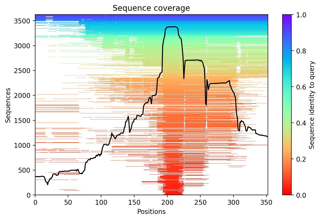
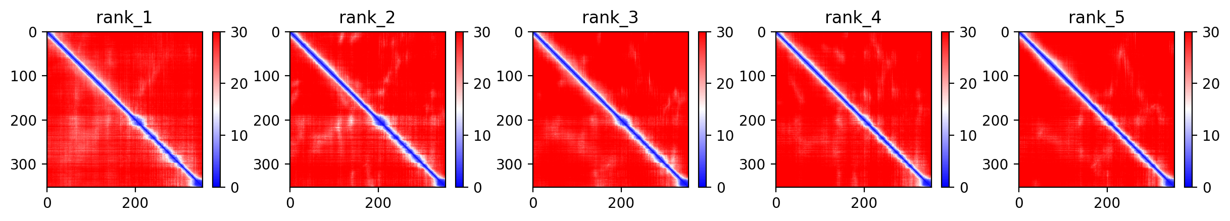
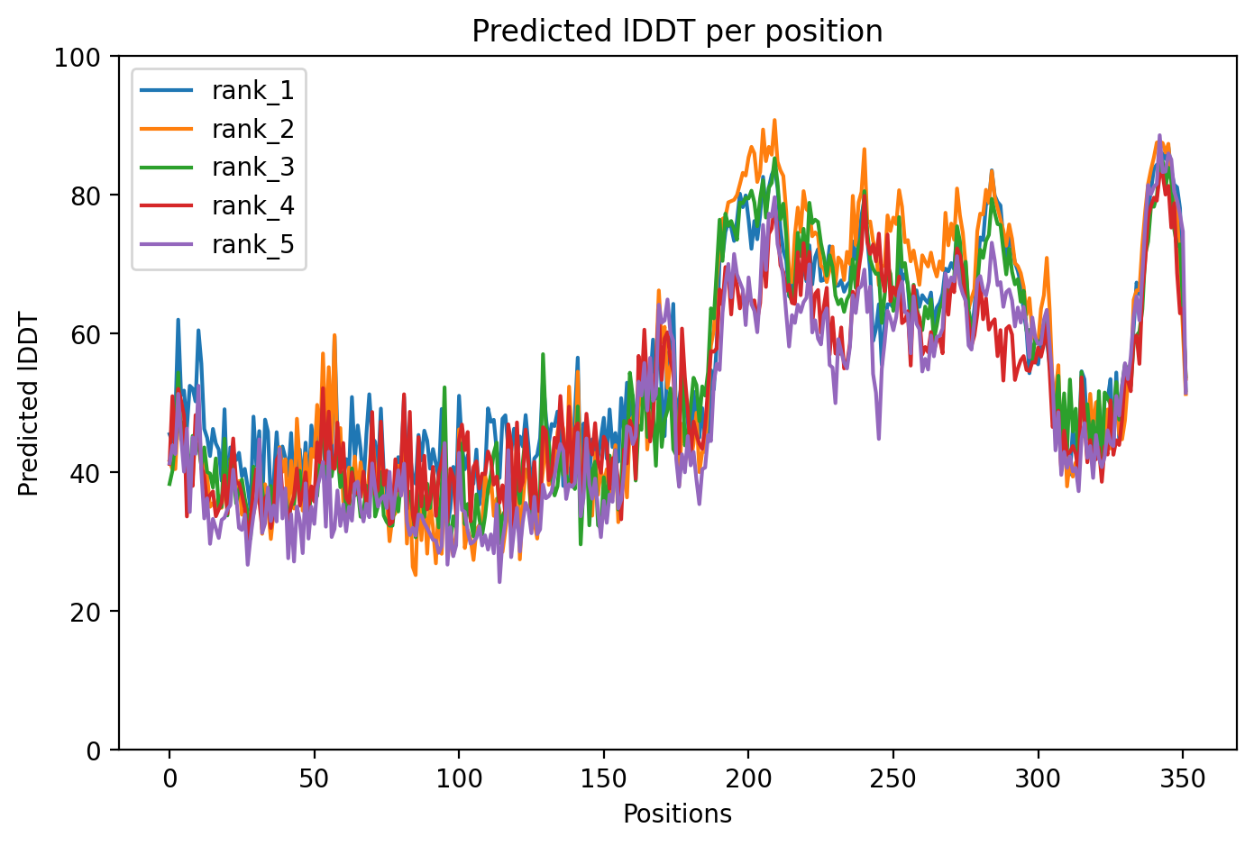
Average pLDDT of 55.0 with only 19% high-confidence residues (67/352) indicates a largely intrinsically disordered protein. The microtubule-binding domain (residues 561-685) shows the highest structural confidence, while N-terminal and C-terminal regions remain destabilized.

The four tandem Tau/MAP repeats (residues 561-685) constitute the microtubule-binding domain and represent the most structured region. Extensive disordered regions (residues 1-573, 715-734) and low complexity segments correlate with low pLDDT throughout most of the protein, consistent with Tau's known intrinsically disordered character.

The P301L mutation in the microtubule-binding domain disrupts proline-mediated structural constraints, potentially accelerating pathological aggregation and reducing microtubule-binding affinity characteristic of frontotemporal dementia-associated Tau variants.

07/Peptide Therapeutics

Aggregation analysis pending. Run peptide agent to compute aggregation propensity.

08/Known Inhibitors

No known inhibitors found. Run peptide agent to search literature.

09/Candidate Peptides

No candidate peptides generated yet. Run peptide agent to design inhibitory peptides.

10/Agent Findings

0 findings

No agent findings yet. Research agents analyze folds on scheduled intervals.

11/Agent Annotations

0 annotations

No agent annotations yet. Agents can submit annotations via the API.