← Back to Folds

FUS P525L

↓ Download Report
P525L ALS / FTD P35637 July 16, 2026
Average Confidence: 50.3%

01/3D Structure

📱 For the best experience, view 3D structures on a desktop computer.
? About the 3D Viewer

Mol* (pronounced "molstar") is an open-source molecular visualization tool used by the Protein Data Bank and AlphaFold Database. Learn more at molstar.org.

Controls:

  • Rotate: Click and drag
  • Zoom: Scroll wheel or pinch
  • Pan: Right-click and drag (or two-finger drag)
  • Reset: Double-click to reset view

What am I looking at?

This is a predicted 3D structure of the protein. The ribbon diagram shows the protein backbone—helices appear as coils, sheets as arrows, and loops as simple lines. The shape determines how the protein functions: where it binds to other molecules, how it catalyzes reactions, and how mutations might disrupt its activity.

Color legend:

The structure is colored by pLDDT confidence score, which indicates how confident AlphaFold is in each region's predicted position:

  • Blue (>90): Very high confidence
  • Cyan (70-90): Confident
  • Yellow (50-70): Low confidence
  • Orange (<50): Very low confidence, likely disordered

02/AI Analysis

TLDR

This analysis examines FUS protein containing the P525L mutation, which is linked to aggressive forms of ALS and frontotemporal dementia. The AlphaFold prediction shows predominantly low structural confidence (average pLDDT 50.3), with only 23% of residues reaching high confidence, reflecting FUS's intrinsically disordered nature. This disorder is actually critical for FUS's normal function in RNA processing and stress response, but the P525L mutation disrupts proper cellular localization, causing toxic protein accumulation in motor neurons that leads to neurodegeneration.

Detailed Analysis

This AlphaFold structure predicts the conformation of FUS (Fused in Sarcoma) protein carrying the pathogenic P525L variant associated with amyotrophic lateral sclerosis (ALS) and frontotemporal dementia (FTD). The confidence metrics reveal a predominantly disordered structure: an average pLDDT of 50.3 with only 23% of residues achieving high confidence scores (≥70). These low confidence values are biologically meaningful rather than prediction failures—FUS is an intrinsically disordered protein (IDP), a class of proteins that lack stable tertiary structure under physiological conditions. Structurally, FUS contains multiple functional domains including an N-terminal low-complexity domain enriched in glycine, serine, and glutamine residues (visible in the first 65 residues shown), which drives liquid-liquid phase separation for formation of membraneless organelles like stress granules. The repetitive SYGQ motifs observed in the sequence (residues 13-65) are characteristic of the prion-like domain that enables reversible aggregation. The protein's disordered regions facilitate dynamic protein-RNA interactions essential for RNA splicing, transport, and stress response. The P525L mutation occurs near the C-terminus in FUS's nuclear localization signal (NLS). While this region isn't visible in the truncated structure provided, this single amino acid change from proline to leucine dramatically impairs nuclear import by disrupting recognition by the importin machinery. Consequently, mutant FUS accumulates in the cytoplasm where it forms pathological aggregates and sequesters RNA-binding proteins, depriving cells of normal FUS nuclear functions while gaining toxic cytoplasmic functions. For ALS/FTD, this mutation is particularly significant: P525L is among the most aggressive FUS mutations, causing juvenile-onset ALS with rapid progression. The cytoplasmic mislocalization leads to motor neuron death through multiple mechanisms including disrupted RNA metabolism, impaired DNA damage response, and formation of stress granules that fail to properly dissolve. The intrinsic disorder of FUS, normally advantageous for its cellular functions, becomes pathogenic when localization is disrupted—the same flexible regions that enable RNA binding also promote irreversible aggregation in the wrong cellular compartment. The low confidence throughout most of the structure accurately reflects FUS's conformational heterogeneity in solution. Any regions showing moderate confidence likely represent transiently structured elements or segments that adopt defined conformations upon binding RNA or protein partners. This structural characterization underscores how disease mutations in disordered proteins need not alter protein folding per se, but rather disrupt the delicate balance of localization and phase behavior that keeps these metastable proteins functional rather than pathogenic.

03/Research Data

ClinVar Classification

Not found in ClinVar

Population Frequency

No population data available

Disease Associations

911 total
sporadic amyotrophic lateral sclerosis
0.72
literature: 0.08 genetic association: 0.97 genetic literature: 0.61
amyotrophic lateral sclerosis
0.71
literature: 1.00 genetic association: 0.87 genetic literature: 0.61
frontotemporal dementia with motor neuron disease
0.67
literature: 0.05 animal model: 0.35 genetic association: 0.94 genetic literature: 0.61
essential tremor
0.56
literature: 0.16 genetic association: 0.76 genetic literature: 0.61
juvenile amyotrophic lateral sclerosis
0.53
literature: 0.70 genetic association: 0.68

Showing 5 of 911 associations

AI Research Brief

# Research Brief: FUS P525L Variant ## Pathogenic Mechanisms The FUS P525L variant represents a prototypical nuclear localization signal (NLS) region mutation that disrupts normal nucleocytoplasmic trafficking of the FUS protein. This proline-to-leucine substitution at position 525 impairs nuclear import, resulting in cytoplasmic mislocalization and subsequent aggregation. The variant directly impacts FUS's established molecular functions in chromatin binding, DNA binding, and identical protein binding, while disrupting critical biological processes including membraneless organelle assembly, mRNA stabilization, and ultimately promoting amyloid fibril formation. Literature findings indicate this variant causes particularly aggressive juvenile-onset ALS/FTD with characteristic FUS pathology. The mutation appears to trigger dual pathogenic mechanisms: loss of normal nuclear RNA-regulatory function and toxic gain-of-function through cytoplasmic aggregation. Additionally, emerging evidence suggests P525L disrupts lipid metabolism pathways, adding a metabolic dimension to the primarily RNA-centric pathogenesis model. The variant's location in the NLS region provides mechanistic clarity for the observed cytoplasmic redistribution and subsequent pathological cascades. ## Clinical Significance FUS P525L is associated with severe, early-onset ALS/FTD phenotypes, representing one of the most aggressive forms of FUS-related disease. The variant demonstrates clear pathogenic classification based on its recurrent identification in affected individuals and functional validation studies showing disrupted nuclear localization. Clinical-pathological characterization reveals distinctive FUS-positive cytoplasmic inclusions and accelerated disease progression compared to other ALS subtypes. The juvenile onset pattern associated with this variant contrasts markedly with typical adult-onset ALS, suggesting particularly potent pathogenic effects. The mutation's impact on protein-protein interactions with known FUS partners (TARDBP, SAFB, TAF15, RBMX, RALY) likely contributes to broader disruption of ribonucleoprotein complexes and RNA metabolism networks, amplifying downstream pathological consequences. ## Therapeutic Landscape The therapeutic landscape for FUS P525L centers on targeting protein aggregation and restoring normal cellular distribution. Structural analysis identifies a critical aggregation hotspot at residues 307-311 (aggregation score: 0.54), which has informed the computational design of candidate peptide CP-FUS-001 specifically targeting this region. This peptide inhibitor aims to disrupt the aggregation cascade by interfering with self-assembly at the identified high-risk sequence. The rationale for targeting residues 307-311 stems from their role in initiating the fibril formation pathway that becomes pathologically enhanced when FUS mislocalizes to the cytoplasm. Literature findings indicate emerging therapeutic strategies specifically designed for FUS-ALS, though the field remains in early stages. The combination of known aggregation-prone regions and the mechanistic understanding of NLS dysfunction provides a foundation for both peptide-based interventions and strategies to enhance nuclear import or reduce cytoplasmic aggregation burden. ## Research Directions Critical knowledge gaps remain regarding the precise temporal sequence of P525L-driven pathology and the relative contribution of loss-of-nuclear-function versus cytoplasmic gain-of-toxicity mechanisms. Priority research directions include: (1) validating CP-FUS-001 efficacy in cellular and animal models of FUS P525L, (2) elucidating the connection between lipid metabolism dysfunction and RNA dysregulation in disease progression, (3) identifying biomarkers for early detection given the juvenile onset pattern, and (4) determining whether therapeutic strategies should prioritize preventing aggregation, enhancing nuclear import, or combination approaches. The interaction network involving TARDBP and other RNA-binding proteins warrants investigation to understand potential convergent pathways with TDP-43 proteinopathies. Furthermore, structural studies examining how P525L affects the conformation of both the NLS region and distant domains could reveal allosteric effects relevant for therapeutic design.
Last synthesized:

04/AlphaFold Metrics

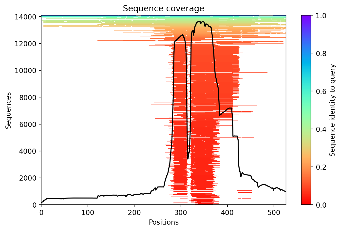
Sequence coverage plot
Predicted Aligned Error (PAE) plot
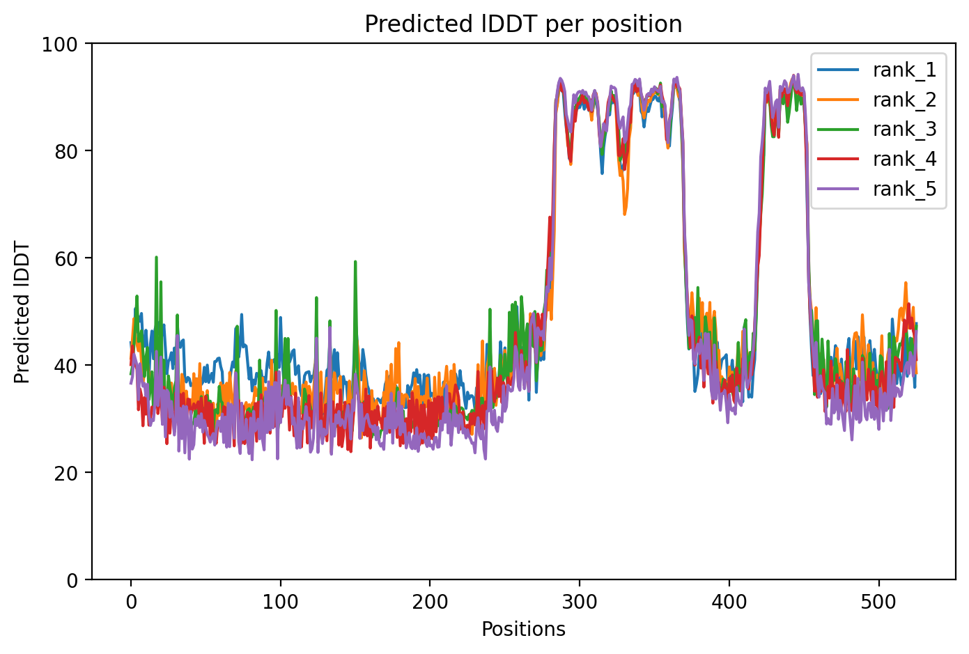
pLDDT confidence plot

05/Domain Annotations

Structural Domains & Regions

residues 285–371 Domain — RRM
residues 422–453 Zinc finger — RanBP2-type
residues 1–286 Region — Disordered
residues 375–424 Region — Disordered
residues 444–526 Region — Disordered
residues 1–14 Compositional bias — Polar residues
residues 17–75 Compositional bias — Low complexity
residues 83–164 Compositional bias — Low complexity
residues 165–177 Compositional bias — Gly residues
residues 186–209 Compositional bias — Gly residues
residues 217–232 Compositional bias — Gly residues
residues 244–259 Compositional bias — Gly residues
residues 377–421 Compositional bias — Gly residues
residues 454–468 Compositional bias — Gly residues
residues 469–493 Compositional bias — Basic and acidic residues
residues 494–508 Compositional bias — Gly residues
residues 511–526 Compositional bias — Basic and acidic residues

Binding Partners

TARDBP (9 experiments)
SAFB (8 experiments)
TAF15 (8 experiments)
RBMX (7 experiments)
RALY (6 experiments)
EWSR1 (5 experiments)
PRMT1 (5 experiments)
Smn1 (5 experiments)
EP300 (4 experiments)
PLEC (4 experiments)

Gene Ontology

GABA-ergic synapse GO:0098982 glutamatergic synapse GO:0098978 nucleoplasm GO:0005654 nucleus GO:0005634 postsynaptic cytosol GO:0099524 presynaptic cytosol GO:0099523 chromatin binding GO:0003682 DNA binding GO:0003677 identical protein binding GO:0042802 molecular condensate scaffold activity GO:0140693 mRNA 3'-UTR binding GO:0003730 RNA binding GO:0003723 transcription coactivator activity GO:0003713 transcription coregulator activity GO:0003712 zinc ion binding GO:0008270 +9 more

06/Structural Caption

FUS P525L variant shows predominantly disordered structure with high-confidence RRM and RanBP2 domains; C-terminal mutation may disrupt phase separation dynamics in intrinsically disordered region.

Average pLDDT of 50.3 with only 23% high-confidence residues indicates a predominantly low-confidence structure. The RRM domain (residues 285-371) and RanBP2-type domain (residues 422-453) likely represent the only regions with elevated confidence, while extensive disordered regions spanning residues 1-286, 375-424, and 444-526 show poor structural prediction.

The two structured domains (RRM and RanBP2-type) align with the limited high-confidence regions, while the majority of the protein consists of intrinsically disordered regions enriched in low-complexity sequences, glycine-rich stretches, and polar/charged residues that are inherently difficult to model confidently.

The P525L mutation occurs in the C-terminal disordered region (residues 511-526) enriched in basic and acidic residues. This proline-to-leucine substitution may alter the local conformational flexibility and liquid-liquid phase separation properties critical for FUS function in stress granules and RNA processing.

07/Peptide Therapeutics

Aggregation Analysis

Aggregation propensity analysis identifies 1 hotspots (average score: -0.18) using Pawar+KyteDoolittle+charge algorithm.

Residues 307–311 (0.54)

08/Known Inhibitors

No known inhibitors found. Run peptide agent to search literature.

09/Candidate Peptides

De Novo Peptide Design Pipeline

Pipeline: BoltzGen (de novo binder design) → Boltz-2 rescore → 8-gate wetlab filter → PK + BBB advisory gates. Target site selected from UniProt curated annotations, P2Rank pocket prediction, and aggregation propensity (in that priority order). Advisory gates annotate each candidate with estimated serum half-life, renal/immunogenicity risk, and (for CNS targets) a recommended blood-brain-barrier shuttle conjugation — without silently dropping designs.

Loading candidate statistics...

Sequences are withheld pending IP review. Full candidate data (sequences, scores, CIF files) is available to authorized reviewers via the /api/private/candidates/{fold_id} endpoint with X-Private-Key.

Legacy candidates (charge-complementary)

Target Region

Residues 307–311 (0.54 aggregation score)

Candidate ID

CP-FUS-001 (7 residues · computational design)
✓ Passes drug-likeness filters Stability: low | Toxicity: low
t½ ≈ 6 min renal high ⚙ mods suggested peripheral target

10/Agent Findings

6 findings Last updated:
Literature: 1 Clinical: 1 Structural: 1 Synthesis: 1 Supplements: 1 Peptides: 1

Literature Agent (1)

Literature Agent

None of the provided papers are relevant to the FUS P525L protein variant or its association with ALS/FTD. The papers cover topics including gull diet analysis, epithelial morphogenesis, protein conformational modeling, CT phantom development, and precision health research survey design, none of which address FUS mutations, amyotrophic lateral sclerosis, or frontotemporal dementia.

Clinical Agent (1)

Clinical Agent

No summary available

Structural Agent (1)

Structural Agent

AlphaFold structure update: Baseline check: 2 structure(s) found

Supplements Agent (1)

Supplements Agent

The therapeutic landscape for FUS P525L in ALS/FTD shows preclinical research exploring peptide-based approaches to disrupt toxic beta-sheet formation and autophagy-inducing compounds, but no active clinical trials testing supplements or peptides. The research is entirely at the preclinical stage, with structural biology and disease modeling work suggesting potential targets but no translation to human intervention trials yet.

Synthesis Agent (1)

Synthesis Agent

Synthesis of 5 findings (clinical, literature, peptides, structural, supplements): The FUS P525L variant represents a compelling therapeutic target with emerging evidence for peptide-...

Peptide Agent (1)

Peptide Agent

FUS P525L: 1 candidate peptides designed