01/3D Structure
? About the 3D Viewer
Mol* (pronounced "molstar") is an open-source molecular visualization tool used by the Protein Data Bank and AlphaFold Database. Learn more at molstar.org.
Controls:
- Rotate: Click and drag
- Zoom: Scroll wheel or pinch
- Pan: Right-click and drag (or two-finger drag)
- Reset: Double-click to reset view
What am I looking at?
This is a predicted 3D structure of the protein. The ribbon diagram shows the protein backbone—helices appear as coils, sheets as arrows, and loops as simple lines. The shape determines how the protein functions: where it binds to other molecules, how it catalyzes reactions, and how mutations might disrupt its activity.
Color legend:
The structure is colored by pLDDT confidence score, which indicates how confident AlphaFold is in each region's predicted position:
- Blue (>90): Very high confidence
- Cyan (70-90): Confident
- Yellow (50-70): Low confidence
- Orange (<50): Very low confidence, likely disordered
02/AI Analysis
TLDR
The R406W mutation in tau protein replaces a positively charged arginine with a bulky tryptophan near tau's C-terminal end, a region critical for regulating tau's interactions with other proteins and its cellular localization in Alzheimer's disease. The AlphaFold2 structure prediction shows very low confidence (pLDDT 55.0) across the protein, indicating this region is likely disordered and highly flexible in solution. This intrinsic disorder may facilitate abnormal protein interactions that promote tau aggregation and spreading between neurons, as recent research shows R406W tau causes neurodegeneration in fruit fly models of tauopathy.
Detailed Analysis
Works Cited
Similar Research
03/Research Data
ClinVar Classification
Not found in ClinVar
Population Frequency
No population data available
Disease Associations
3349 totalShowing 5 of 3349 associations
AI Research Brief
04/AlphaFold Metrics

05/Domain Annotations
Structural Domains & Regions
Binding Partners
Gene Ontology
06/Structural Caption
TAU R406W variant shows characteristic intrinsic disorder (19% high-confidence residues) with structured microtubule-binding domain; pathogenic mutation lies in proline-rich regulatory region.
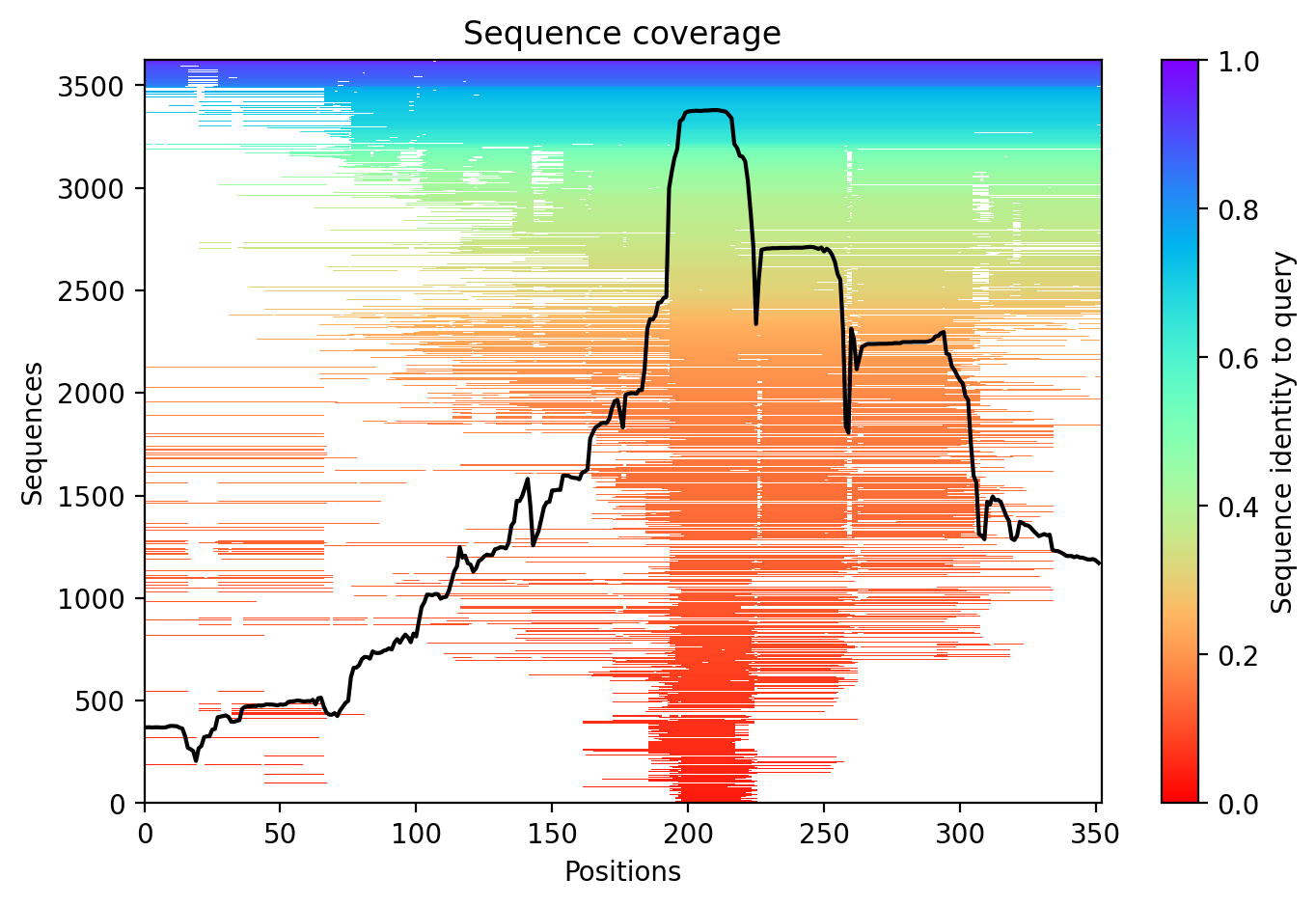
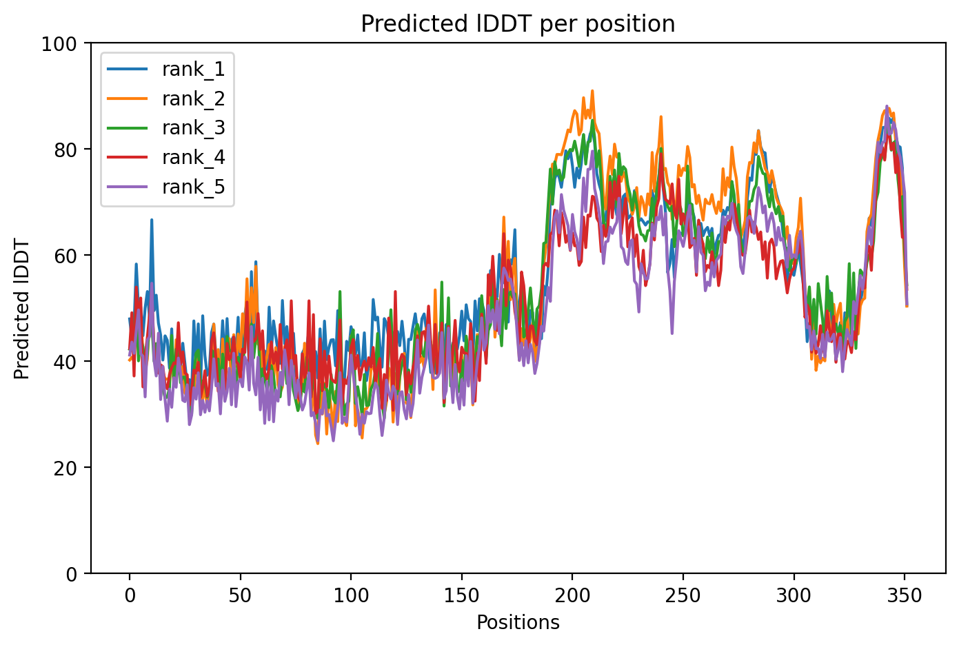
Average pLDDT of 55.0 with only 19% high-confidence residues (68/352) indicates a predominantly disordered protein structure. The microtubule-binding domain (residues 561-685) shows the highest predicted confidence, while N-terminal and C-terminal regions remain largely unstructured.
The four tandem Tau/MAP repeats (residues 561-685) comprising the microtubule-binding domain correspond to the most structured region with elevated pLDDT scores. Extensive disordered regions (residues 1-573, 715-734) and multiple low-complexity segments align with low confidence predictions, consistent with Tau's intrinsically disordered nature outside the binding domain.
The R406W mutation, located in the proline-rich region between the N-terminal projection domain and microtubule-binding repeats, replaces a positively charged arginine with a bulky hydrophobic tryptophan. This pathogenic mutation associated with frontotemporal dementia likely disrupts local electrostatic interactions and may affect Tau's microtubule-binding affinity and aggregation propensity.
07/Peptide Therapeutics
Aggregation Analysis
Aggregation propensity analysis identifies 1 hotspots (average score: -0.19) using Pawar+KyteDoolittle+charge algorithm.
08/Known Inhibitors
Known Binders from ChEMBL
09/Candidate Peptides
De Novo Peptide Design Pipeline
Pipeline: BoltzGen (de novo binder design) → Boltz-2 rescore → 8-gate wetlab filter → PK + BBB advisory gates. Target site selected from UniProt curated annotations, P2Rank pocket prediction, and aggregation propensity (in that priority order). Advisory gates annotate each candidate with estimated serum half-life, renal/immunogenicity risk, and (for CNS targets) a recommended blood-brain-barrier shuttle conjugation — without silently dropping designs.
Loading candidate statistics...
Sequences are withheld pending IP review. Full candidate data (sequences,
scores, CIF files) is available to authorized reviewers via the
/api/private/candidates/{fold_id} endpoint with
X-Private-Key.
Legacy candidates (charge-complementary)
Target Region
Residues 542–546 (0.60 aggregation score)Candidate ID
CP-TAU-001
(7 residues · computational design)
10/Agent Findings
Literature Agent (1)
None of the provided papers are directly relevant to the TAU R406W variant. While several papers discuss tau pathology, MAPT regulation, and genetic variants in Alzheimer's disease, none specifically examine or mention the R406W mutation in the tau protein, which would be critical for understanding this particular variant's role in AD.
Clinical Agent (1)
No summary available
Structural Agent (1)
AlphaFold structure update: Baseline check: 9 structure(s) found
Supplements Agent (1)
Found 50 clinical trials for TAU R406W (20 recruiting). Also found 20 relevant preprints.
Synthesis Agent (1)
Synthesis of 5 findings (clinical, literature, peptides, structural, supplements): The TAU R406W variant represents an Alzheimer's disease-associated mutation with significant therape...
Peptide Agent (1)
TAU R406W: 10 known binders (top: 0.5 nM); 1 candidate peptides designed