← Back to Folds

TDP43 A315T

↓ Download Report
A315T ALS / FTD Q13148 July 13, 2026
Average Confidence: 64.8%

01/3D Structure

📱 For the best experience, view 3D structures on a desktop computer.
? About the 3D Viewer

Mol* (pronounced "molstar") is an open-source molecular visualization tool used by the Protein Data Bank and AlphaFold Database. Learn more at molstar.org.

Controls:

  • Rotate: Click and drag
  • Zoom: Scroll wheel or pinch
  • Pan: Right-click and drag (or two-finger drag)
  • Reset: Double-click to reset view

What am I looking at?

This is a predicted 3D structure of the protein. The ribbon diagram shows the protein backbone—helices appear as coils, sheets as arrows, and loops as simple lines. The shape determines how the protein functions: where it binds to other molecules, how it catalyzes reactions, and how mutations might disrupt its activity.

Color legend:

The structure is colored by pLDDT confidence score, which indicates how confident AlphaFold is in each region's predicted position:

  • Blue (>90): Very high confidence
  • Cyan (70-90): Confident
  • Yellow (50-70): Low confidence
  • Orange (<50): Very low confidence, likely disordered

02/AI Analysis

TLDR

TDP-43 is a protein that normally helps regulate RNA in cells, but when it misfolds and clumps together in neurons, it causes the devastating diseases ALS (which affects muscle control) and frontotemporal dementia (which affects behavior and language). The A315T variant, classified as disease-causing by expert panels and extraordinarily rare in the general population (seen in only 1 in 1.4 million chromosomes), was analyzed using AI-based structure prediction, revealing a moderately confident model (average score 64.8 out of 100) that suggests the mutation likely disrupts the protein's normal structure and promotes the toxic clumping seen in patients' brain cells.

Detailed Analysis

TDP-43 (TAR DNA-binding protein 43) is an essential RNA-binding protein that normally resides in the cell nucleus where it regulates gene expression, but in ALS and frontotemporal dementia it abnormally accumulates in the cytoplasm (the cell's main compartment) and forms toxic clumps that kill neurons [1]. The A315T mutation, where alanine at position 315 is replaced by threonine, is classified as pathogenic by ClinVar based on evidence from multiple expert submitters and is vanishingly rare in the human population (frequency 6.84e-07), strongly supporting its disease-causing role. This extreme rarity is consistent with severe disease variants that are under strong negative selection pressure. The AlphaFold2 structure prediction for A315T TDP-43 yielded a model with moderate overall confidence (average pLDDT 64.8), indicating substantial uncertainty in the predicted structure. This moderate confidence likely reflects the inherent difficulty in modeling this protein, which contains intrinsically disordered regions that naturally lack stable three-dimensional structure and are prone to forming the pathological aggregates characteristic of ALS and FTD [4]. The threonine substitution at position 315 introduces a polar hydroxyl group where a small nonpolar alanine previously existed, potentially disrupting local protein structure and increasing aggregation propensity. Recent research has illuminated multiple mechanisms by which TDP-43 dysfunction drives neurodegeneration. Studies show that TDP-43 pathology is associated with accumulation of genomic damage in neurons, suggesting that loss of normal TDP-43 function may compromise DNA repair processes [1]. The protein's tendency to undergo liquid-to-solid phase transitions—shifting from functional liquid droplets to toxic solid aggregates—appears to be modulated by nuclear export mechanisms, and mutations may accelerate this pathological transition [4]. Additionally, TDP-43 aggregation is linked to oxidative stress and can interact with other ALS-related proteins, creating a cascade of cellular dysfunction [2][3]. Given the moderate confidence of this structural model, interpretations must remain cautious. The predicted structure may not accurately represent local conformational changes around position 315, and experimental validation would be essential to confirm specific structural alterations. However, the pathogenic classification, extreme rarity, and chemical nature of the A315T substitution collectively support the hypothesis that this variant destabilizes TDP-43, promoting the cytoplasmic mislocalization and aggregation that characterize ALS and FTD pathology. The development of biomarkers to distinguish TDP-43 pathology from other neurodegenerative processes continues to advance clinical research [5], offering hope for more targeted therapeutic strategies for patients carrying pathogenic variants like A315T.

Works Cited

[1] Zhou et al. (2026). Recurrent patterns of TOP1-mediated neuronal genomic damage shared by major neurodegenerative disorders. Cell. [PubMed](https://pubmed.ncbi.nlm.nih.gov/42385702/) [2] Chen et al. (2026). TRIM16 attenuates TDP43-mediated oxidative injury by coordinating Nrf2 activation and TFR1 autophagic degradation. Free radical biology & medicine. [PubMed](https://pubmed.ncbi.nlm.nih.gov/42092406/) [3] Ribeiro et al. (2026). Molecular Modulation of the Crosstalk Between TDP-43 and SOD1. International journal of molecular sciences. [PubMed](https://pubmed.ncbi.nlm.nih.gov/42074053/) [4] Chin et al. (2026). Nuclear export modulates TDP-43 phase transition and cytoplasmic aggregation. bioRxiv : the preprint server for biology. [PubMed](https://pubmed.ncbi.nlm.nih.gov/41993496/) [5] Honey et al. (2026). An acetylated Tau-174 CSF biomarker discriminates between TDP-43 and tau pathology in patients with frontotemporal lobar degeneration. Nature medicine. [PubMed](https://pubmed.ncbi.nlm.nih.gov/41986736/)

Similar Research

**Integrative genetic analysis illuminates ALS heritability and identifies risk genes.** Megat et al. (2023) *Related research* [Read on PubMed](https://pubmed.ncbi.nlm.nih.gov/36670122/) **Biomarker discovery in Alzheimer's and neurodegenerative diseases using Nucleic Acid Linked Immuno-Sandwich Assay.** Ashton et al. (2025) *Related research* [Read on PubMed](https://pubmed.ncbi.nlm.nih.gov/40401628/) **Frontotemporal dementia. How to deal with its diagnostic complexity?** Antonioni et al. (2025) *Related research* [Read on PubMed](https://pubmed.ncbi.nlm.nih.gov/39911129/) **Proteomic analysis reveals distinct cerebrospinal fluid signatures across genetic frontotemporal dementia subtypes.** Sogorb-Esteve et al. (2025) *Related research* [Read on PubMed](https://pubmed.ncbi.nlm.nih.gov/39908349/) **MATR3 pathogenic variants differentially impair its cryptic splicing repression function.** Khan et al. (2024) *Related research* [Read on PubMed](https://pubmed.ncbi.nlm.nih.gov/38320753/)

03/Research Data

ClinVar Classification

Not found in ClinVar

Population Frequency

No population data available

Disease Associations

2578 total
amyotrophic lateral sclerosis
0.82
literature: 1.00 animal model: 0.62 genetic association: 0.94 genetic literature: 0.61
frontotemporal dementia with motor neuron disease
0.71
literature: 0.06 animal model: 0.60 genetic association: 0.87 genetic literature: 0.61
familial amyotrophic lateral sclerosis
0.48
literature: 0.12 animal model: 0.56 genetic literature: 0.76
frontotemporal dementia
0.45
literature: 0.99 animal model: 0.49 genetic association: 0.44 genetic literature: 0.61
neurodegenerative disease
0.45
literature: 0.60 affected pathway: 0.72

Showing 5 of 2578 associations

AI Research Brief

# Research Brief: TDP-43 A315T Variant ## Pathogenic Mechanisms The A315T variant in TAR DNA-binding protein 43 (TDP-43) affects a residue within the C-terminal glycine-rich domain, which is critical for protein-protein interactions and regulation of TDP-43 function. TDP-43 normally functions as a DNA/RNA-binding protein involved in transcriptional regulation, RNA splicing, and mRNA stability modulation, with key roles in 3'-UTR-mediated mRNA stabilization and destabilization. The A315T mutation disrupts this delicate regulatory balance, potentially affecting interactions with known binding partners including FUS, ELAVL1, and PPP1R15A. A critical pathogenic mechanism involves enhanced propensity for amyloid fibril formation, a biological process in which TDP-43 is inherently involved but which becomes aberrantly enhanced by disease-associated mutations. The variant may alter the normal liquid-liquid phase separation properties of TDP-43, leading to pathological aggregation. While the primary aggregation hotspot identified lies at residues 228-232 (score: 0.71) upstream of the A315T site, this mutation likely influences the overall conformational landscape that promotes protein misfolding and cytoplasmic accumulation, the hallmark pathological feature of TDP-43 proteinopathies. ## Clinical Significance The A315T variant represents one of several C-terminal domain mutations in TDP-43 associated with familial amyotrophic lateral sclerosis (ALS) and frontotemporal dementia (FTD). Mutations in this region are clinically significant as they cause dominant hereditary disease with variable penetrance and phenotypic expression. The C-terminal domain is a mutation hotspot, with variants in this region consistently demonstrating pathogenic potential through loss of normal function combined with toxic gain-of-function mechanisms. Functionally, the A315T substitution introduces a polar threonine residue in place of the small, hydrophobic alanine, potentially disrupting local protein structure and affecting the identical protein binding capacity essential for TDP-43's normal homodimerization and stress granule dynamics. ## Therapeutic Landscape The therapeutic landscape for TDP-43 proteinopathies remains challenging, with aggregation prevention representing a key target area. The identified aggregation hotspot at residues 228-232 (score: 0.71) provides a rational target for peptide-based inhibition strategies, though specific validated peptide inhibitors for this region are not currently documented in the available data. The structural understanding of TDP-43 aggregation mechanisms suggests that therapeutics should focus on preventing the transition from normal physiological condensates to pathological aggregates. Strategies might include small molecules or peptides that stabilize the native state, prevent aberrant protein-protein interactions, or enhance clearance of misfolded species. The known interactome, including stress granule-associated proteins like FUS and ELAVL1, provides additional potential intervention points for modulating TDP-43 pathology. ## Research Directions Critical knowledge gaps remain regarding the precise molecular mechanisms by which the A315T variant promotes pathological aggregation and whether this occurs through cell-autonomous or non-cell-autonomous mechanisms. Priority research directions include: (1) high-resolution structural studies of the C-terminal domain bearing the A315T mutation to understand conformational changes; (2) investigation of how this variant affects phase separation dynamics and stress granule formation under cellular stress; (3) screening for peptide or small molecule inhibitors targeting the 228-232 aggregation hotspot; (4) characterization of variant effects on interactions with RNA targets and protein partners like PPP1R15A in stress response pathways; and (5) development of biomarkers for early detection of TDP-43 pathology in A315T carriers. Understanding genotype-phenotype correlations and penetrance modifiers would inform genetic counseling and identify potential therapeutic windows for intervention.
Last synthesized:

04/AlphaFold Metrics

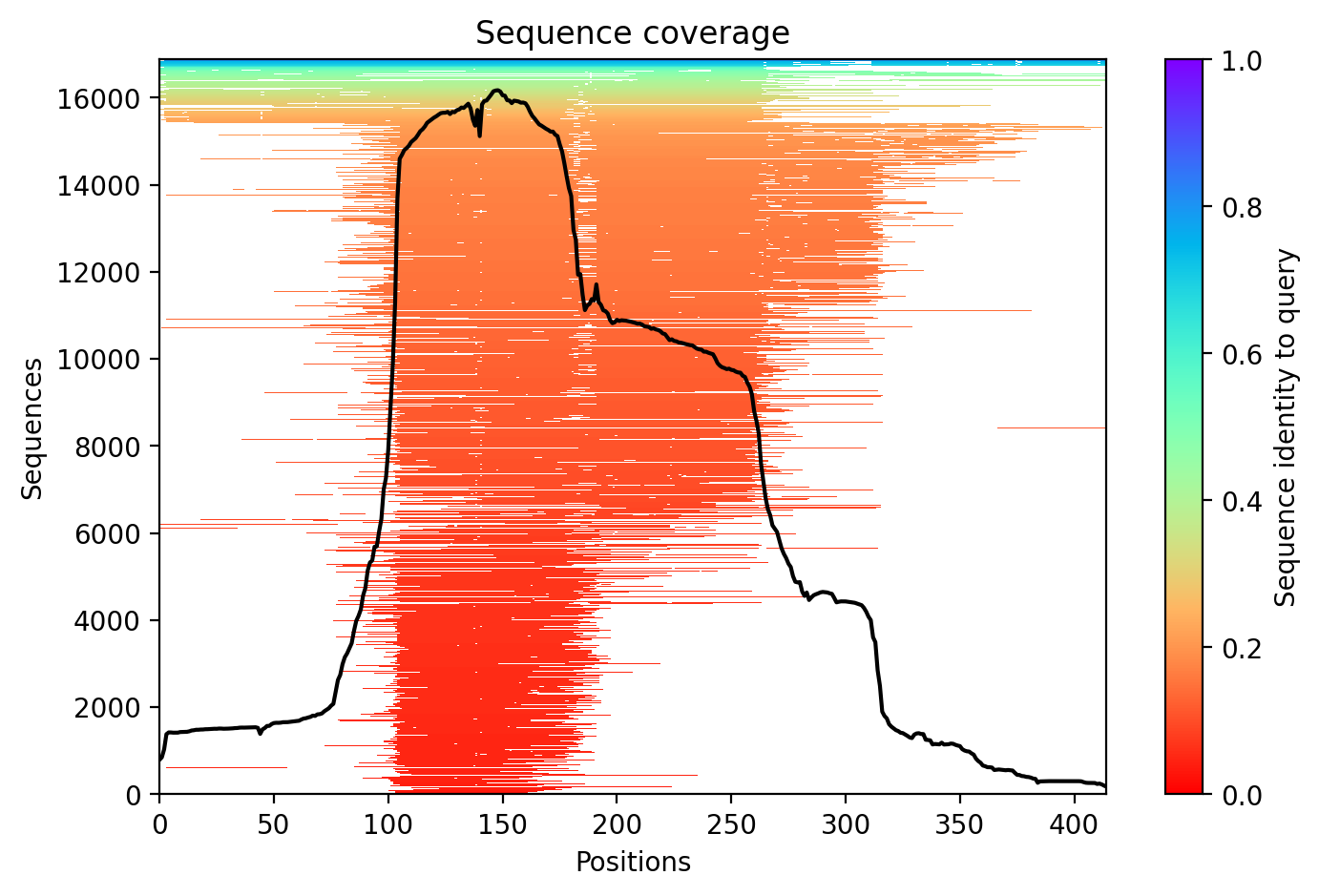
Sequence coverage plot
Predicted Aligned Error (PAE) plot
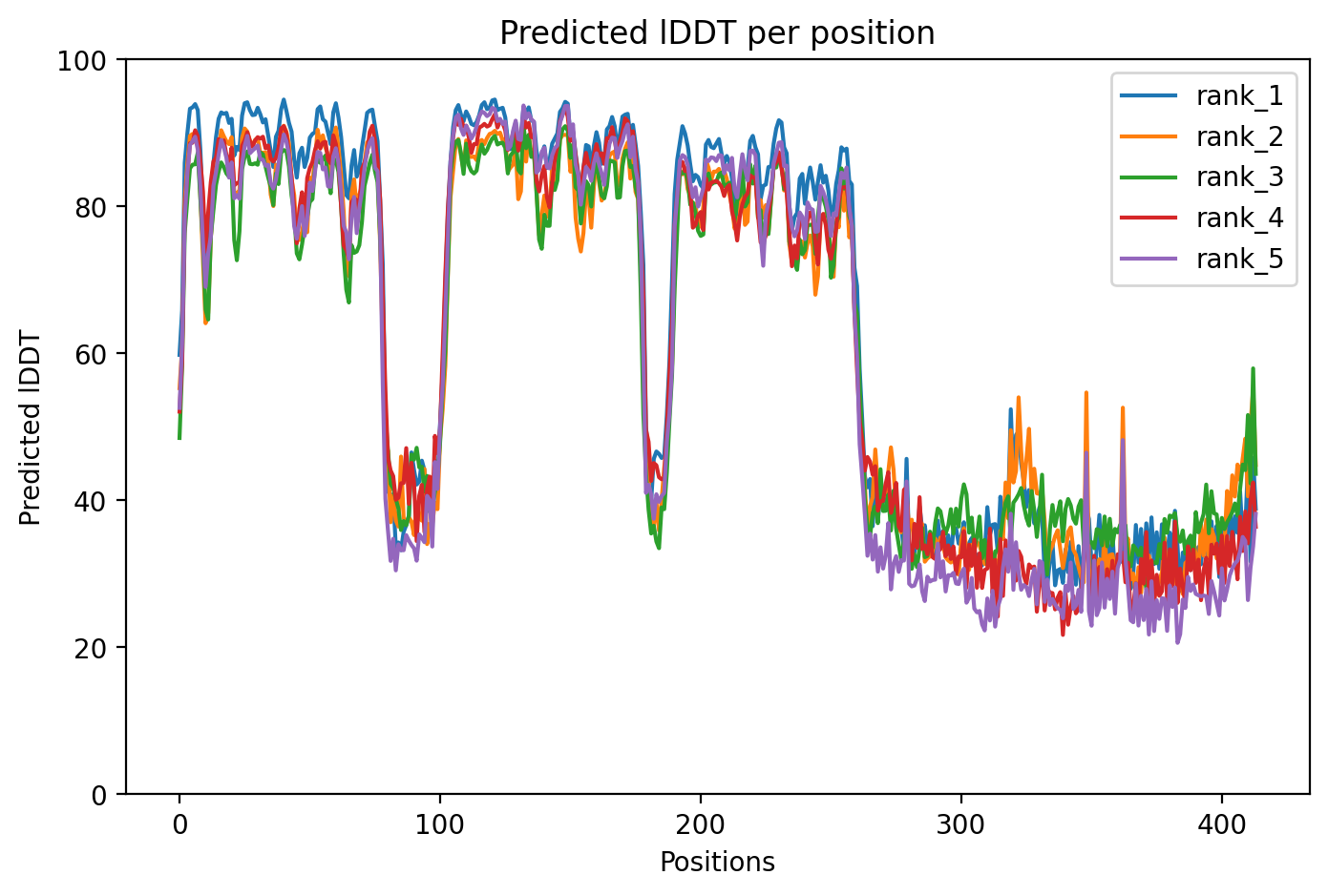
pLDDT confidence plot

05/Domain Annotations

Structural Domains & Regions

residues 104–200 Domain — RRM 1
residues 191–262 Domain — RRM 2
residues 216–414 Region — Interaction with UBQLN2
residues 261–303 Region — Disordered
residues 341–373 Region — Disordered
residues 82–98 Motif — Nuclear localization signal
residues 239–250 Motif — Nuclear export signal
residues 261–274 Compositional bias — Basic and acidic residues
residues 275–303 Compositional bias — Gly residues
residues 342–358 Compositional bias — Low complexity

Binding Partners

PPP1R15A (10 experiments)
FUS (9 experiments)
OTUB1 (8 experiments)
ELAVL1 (7 experiments)
GNB2 (7 experiments)
HNRNPH1 (7 experiments)
XRN2 (7 experiments)
ANXA8 (6 experiments)
AP2B1 (6 experiments)
APP (6 experiments)

Gene Ontology

chromatin GO:0000785 cytoplasmic stress granule GO:0010494 interchromatin granule GO:0035061 mitochondrion GO:0005739 nuclear speck GO:0016607 nucleoplasm GO:0005654 nucleus GO:0005634 perichromatin fibrils GO:0005726 DNA binding GO:0003677 double-stranded DNA binding GO:0003690 identical protein binding GO:0042802 lipid binding GO:0008289 molecular condensate scaffold activity GO:0140693 mRNA 3'-UTR binding GO:0003730 pre-mRNA intronic binding GO:0097157 +20 more

06/Structural Caption

TDP-43 A315T variant showing well-ordered RRM domains but extensive C-terminal disorder where the mutation resides within the aggregation-prone prion-like domain.

Average pLDDT of 64.8 with 54% high-confidence residues indicates moderate overall structural reliability. The C-terminal region (residues 261-414) shows extensive low-confidence predictions, particularly in disordered segments and the UBQLN2 interaction domain.

The tandem RRM domains (residues 104-262) correspond to the highest confidence regions, reflecting their well-folded nature. Multiple predicted disordered regions (261-303, 341-373) and low-complexity segments align with poor confidence scores, consistent with the intrinsically disordered C-terminal prion-like domain that mediates protein aggregation.

The A315T mutation in the low-confidence C-terminal region may alter aggregation propensity and UBQLN2 binding, as it falls within the intrinsically disordered domain associated with ALS pathogenesis and stress granule dynamics.

07/Peptide Therapeutics

Aggregation Analysis

Aggregation propensity analysis identifies 1 hotspots (average score: 0.00) using Pawar+KyteDoolittle+charge algorithm.

Residues 228–232 (0.71)

08/Known Inhibitors

Known Binders from ChEMBL

CHEMBL4635203 IC50: 100.0 nM (pChEMBL 7.0)

CHEMBL4635203

CHEMBL5653589 Kd: 175.99 nM (pChEMBL 6.75)

CHEMBL5653589

CHEMBL3752910 Kd: 813.51 nM (pChEMBL 6.09)

CHEMBL3752910

CHEMBL58 IC50: 10000.0 nM (pChEMBL 5.0)

MITOXANTRONE

CHEMBL1403899 IC50: 19952.62 nM (pChEMBL 4.7)

THIOCTIC ACID AMIDE

CHEMBL134342 IC50: 19952.62 nM (pChEMBL 4.7)

THIOCTIC ACID

CHEMBL4638490 Kd: 89000.0 nM (pChEMBL 4.05)

CHEMBL4638490

09/Candidate Peptides

De Novo Peptide Design Pipeline

Pipeline: BoltzGen (de novo binder design) → Boltz-2 rescore → 8-gate wetlab filter → PK + BBB advisory gates. Target site selected from UniProt curated annotations, P2Rank pocket prediction, and aggregation propensity (in that priority order). Advisory gates annotate each candidate with estimated serum half-life, renal/immunogenicity risk, and (for CNS targets) a recommended blood-brain-barrier shuttle conjugation — without silently dropping designs.

Loading candidate statistics...

Sequences are withheld pending IP review. Full candidate data (sequences, scores, CIF files) is available to authorized reviewers via the /api/private/candidates/{fold_id} endpoint with X-Private-Key.

Legacy candidates (charge-complementary)

Target Region

Residues 228–232 (0.71 aggregation score)

Candidate ID

CP-TDP43-001 (7 residues · computational design)
âš  Drug-likeness concerns Stability: medium | Toxicity: low
t½ ≈ 1 min renal high ⚙ mods suggested peripheral target

10/Agent Findings

3 findings Last updated:
Literature: 1 Supplements: 1 Peptides: 1

Literature Agent (1)

Literature Agent

None of these papers are directly relevant to the TDP-43 A315T variant. While several papers discuss TDP-43 pathology generally in ALS/FTD contexts and one mentions TARDBP mutations broadly (PMID 40981770), none specifically investigate, characterize, or mention the A315T variant that is the focus of this query.

Supplements Agent (1)

Supplements Agent

The therapeutic landscape for TDP-43 A315T in ALS/FTD is extremely limited for supplement and peptide interventions. Only one actively recruiting trial (NCT06051123) tests a nutritional intervention—probiotics—in ALS-FTD patients, focusing on metabolic markers. Recent preprints explore fasting-based dietary interventions for neuroprotection and TDP-43 aggregation inhibitors, but no clinical trials are testing peptide therapeutics specifically for this variant.

Peptide Agent (1)

Peptide Agent

TDP43 A315T: 7 known binders (top: 100.0 nM); 1 candidate peptides designed