01/3D Structure
? About the 3D Viewer
Mol* (pronounced "molstar") is an open-source molecular visualization tool used by the Protein Data Bank and AlphaFold Database. Learn more at molstar.org.
Controls:
- Rotate: Click and drag
- Zoom: Scroll wheel or pinch
- Pan: Right-click and drag (or two-finger drag)
- Reset: Double-click to reset view
What am I looking at?
This is a predicted 3D structure of the protein. The ribbon diagram shows the protein backbone—helices appear as coils, sheets as arrows, and loops as simple lines. The shape determines how the protein functions: where it binds to other molecules, how it catalyzes reactions, and how mutations might disrupt its activity.
Color legend:
The structure is colored by pLDDT confidence score, which indicates how confident AlphaFold is in each region's predicted position:
- Blue (>90): Very high confidence
- Cyan (70-90): Confident
- Yellow (50-70): Low confidence
- Orange (<50): Very low confidence, likely disordered
02/AI Analysis
TLDR
Alpha-synuclein is a protein whose clumping in brain cells causes Parkinson's disease, and the A30P mutation makes this clumping more likely to occur. Scientists used AlphaFold2 computer modeling to predict the structure of this mutant protein, but the very low confidence score (54% average) indicates the protein lacks stable shape and exists as a disordered molecule. This structural flexibility may explain why A30P alpha-synuclein is particularly prone to forming the toxic clumps that kill dopamine-producing neurons in Parkinson's patients.
Detailed Analysis
Works Cited
Similar Research
03/Research Data
ClinVar Classification
Not found in ClinVar
Population Frequency
No population data available
Disease Associations
801 totalShowing 5 of 801 associations
AI Research Brief
04/AlphaFold Metrics

05/Domain Annotations
Structural Domains & Regions
Functional Sites
Binding Partners
Gene Ontology
06/Structural Caption
Alpha-synuclein A30P variant shows complete structural disorder (0% high-confidence residues), with the pathogenic mutation further destabilizing the N-terminal repeat region implicated in membrane binding.
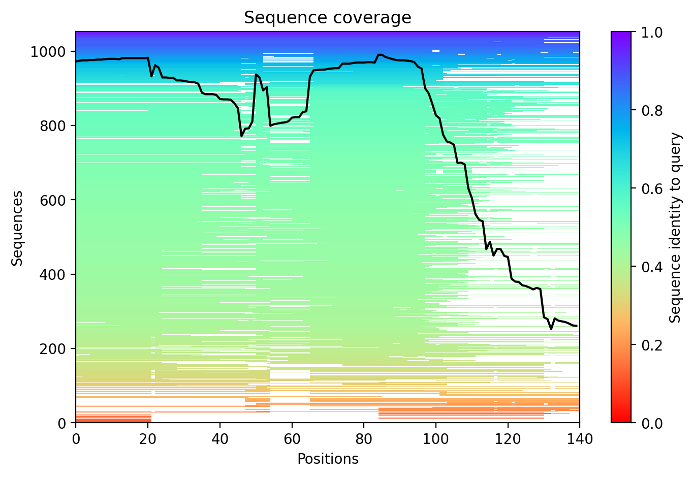
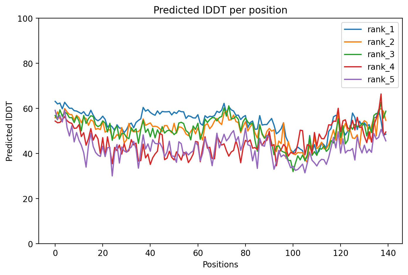
Average pLDDT of 53.8 with 0% high-confidence residues indicates a predominantly disordered structure across all 140 residues. The entire protein shows low structural confidence, consistent with an intrinsically disordered protein lacking stable tertiary structure.
The tandem repeat region (residues 20-67) and C-terminal disordered region (residues 100-140) both show uniformly low confidence, supporting intrinsic disorder rather than stable folding. The SERF1A interaction region (111-140) and acidic C-terminus lack predicted structural stability in isolation.
The A30P mutation in the first tandem repeat region disrupts the already weak structural propensity of alpha-synuclein, likely reducing residual helical tendency and potentially accelerating aggregation. This pathogenic variant associated with early-onset Parkinson's disease destabilizes native conformational ensemble.
07/Peptide Therapeutics
Aggregation Analysis
Aggregation propensity analysis identifies 1 hotspots (average score: 0.00) using Pawar+KyteDoolittle+charge algorithm.
08/Known Inhibitors
Known Binders from ChEMBL
09/Candidate Peptides
De Novo Peptide Design Pipeline
Pipeline: BoltzGen (de novo binder design) → Boltz-2 rescore → 8-gate wetlab filter → PK + BBB advisory gates. Target site selected from UniProt curated annotations, P2Rank pocket prediction, and aggregation propensity (in that priority order). Advisory gates annotate each candidate with estimated serum half-life, renal/immunogenicity risk, and (for CNS targets) a recommended blood-brain-barrier shuttle conjugation — without silently dropping designs.
Loading candidate statistics...
Sequences are withheld pending IP review. Full candidate data (sequences,
scores, CIF files) is available to authorized reviewers via the
/api/private/candidates/{fold_id} endpoint with
X-Private-Key.
Legacy candidates (charge-complementary)
Target Region
Residues 15–19 (0.51 aggregation score)Candidate ID
CP-ALPHA-001
(7 residues · computational design)
10/Agent Findings
Literature Agent (1)
None of the provided papers directly investigated the A30P alpha-synuclein variant. While several papers examined other SNCA mutations (A53T, H50Q) and provided valuable insights into mutation-specific pathogenic mechanisms, fibril polymorphism, and disease progression patterns that may inform understanding of familial PD variants generally, they do not provide specific evidence about A30P pathobiology.
Clinical Agent (1)
No summary available
Structural Agent (1)
AlphaFold structure update: Baseline check: 3 structure(s) found
Supplements Agent (1)
Found 28 clinical trials for ALPHA-SYNUCLEIN A30P (20 recruiting). Also found 15 relevant preprints.
Synthesis Agent (1)
Synthesis of 5 findings (clinical, literature, peptides, structural, supplements): The ALPHA-SYNUCLEIN A30P variant shows a research landscape characterized by indirect therapeutic ap...
Peptide Agent (1)
ALPHA-SYNUCLEIN A30P: 10 known binders (top: 2.1 nM); 1 candidate peptides designed