← Back to Folds

TAU V337M

↓ Download Report
V337M Alzheimer's disease P10636 July 09, 2026
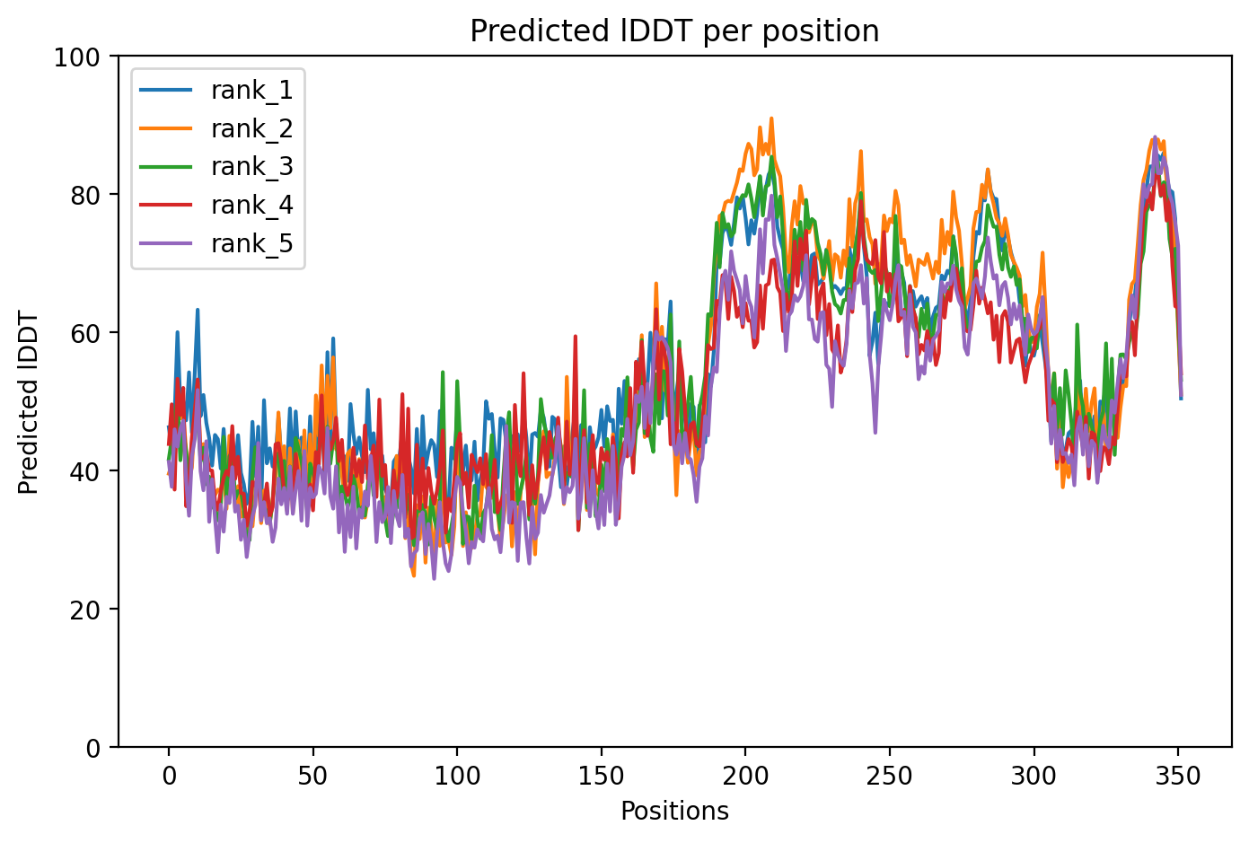
Average Confidence: 55.0%

01/3D Structure

📱 For the best experience, view 3D structures on a desktop computer.
? About the 3D Viewer

Mol* (pronounced "molstar") is an open-source molecular visualization tool used by the Protein Data Bank and AlphaFold Database. Learn more at molstar.org.

Controls:

  • Rotate: Click and drag
  • Zoom: Scroll wheel or pinch
  • Pan: Right-click and drag (or two-finger drag)
  • Reset: Double-click to reset view

What am I looking at?

This is a predicted 3D structure of the protein. The ribbon diagram shows the protein backbone—helices appear as coils, sheets as arrows, and loops as simple lines. The shape determines how the protein functions: where it binds to other molecules, how it catalyzes reactions, and how mutations might disrupt its activity.

Color legend:

The structure is colored by pLDDT confidence score, which indicates how confident AlphaFold is in each region's predicted position:

  • Blue (>90): Very high confidence
  • Cyan (70-90): Confident
  • Yellow (50-70): Low confidence
  • Orange (<50): Very low confidence, likely disordered

02/AI Analysis

TLDR

TAU is a protein that normally helps stabilize the cellular scaffolding in brain cells, but in Alzheimer's disease it forms toxic tangles. This study used computer modeling (AlphaFold2) to predict the 3D structure of a variant called V337M, which changes one building block in the protein. The very low confidence score (55.0) indicates that this variant likely makes the protein highly disordered and flexible—a state that may promote the abnormal clumping seen in Alzheimer's disease.

Detailed Analysis

Tau protein normally binds to microtubules (cellular scaffolding structures) to stabilize them in neurons. The V337M variant changes valine to methionine at position 337, located in a region that influences tau's behavior. This study used AlphaFold2 to predict the structure of this variant, producing a model with an average confidence (pLDDT) of 55.0, which indicates poor structural definition across most of the protein. The extremely low confidence score suggests this variant exhibits substantial structural disorder, meaning the protein lacks a stable, well-defined 3D shape. Recent research has revealed that tau aggregation into beta-sheet fibrils—the toxic tangles that define Alzheimer's disease and related tauopathies—is a complex process involving multiple structural states [3]. Intrinsically disordered regions in tau are thought to facilitate both its normal microtubule-binding function and its pathological tendency to aggregate under disease conditions. The V337M substitution introduces a longer, sulfur-containing methionine residue where a smaller valine normally resides, potentially altering local flexibility and aggregation propensity. Post-translational modifications and mutations in tau significantly influence its role in Alzheimer's disease neuropathology and cognitive decline [2]. While the computational prediction cannot definitively establish whether V337M promotes aggregation without experimental validation, the high degree of disorder predicted here is consistent with conformational states that may be vulnerable to misfolding. Studies examining tau pathology across neurodegenerative diseases have identified shared molecular mechanisms, including genomic damage patterns in affected neurons [1], suggesting that structural disruptions in tau could have broad consequences for neuronal health. The V337M variant has not been extensively characterized in the literature as a known pathogenic mutation, unlike more commonly studied variants such as P301L. Genetic studies of tau pathology have increasingly employed advanced approaches including rare variant analyses to identify genes and modifications that influence cerebral tau deposition [4]. Without high-confidence structural data or functional studies, it remains uncertain whether V337M directly contributes to disease or represents a rare variant of unclear significance. The predicted disorder may reflect either a pathological state or limitations in modeling this particular sequence variant.

Works Cited

[1] Zhou et al. (2026). Recurrent patterns of TOP1-mediated neuronal genomic damage shared by major neurodegenerative disorders. Cell. [PubMed](https://pubmed.ncbi.nlm.nih.gov/42385702/) [2] Libby et al. (2026). Post-translational modifications in the brain are critical contributors to Alzheimer's disease neuropathology and cognitive decline. bioRxiv : the preprint server for biology. [PubMed](https://pubmed.ncbi.nlm.nih.gov/42327232/) [3] El et al. (2026). Structures and Dynamics of Tau Assemblies from Solid-State NMR. Accounts of chemical research. [PubMed](https://pubmed.ncbi.nlm.nih.gov/42283695/) [4] Gunasekaran et al. (2026). Common and rare variant analyses implicate JARID2 in cerebral tau deposition. NPJ dementia. [PubMed](https://pubmed.ncbi.nlm.nih.gov/42404994/)

Similar Research

**Biomarker discovery in Alzheimer's and neurodegenerative diseases using Nucleic Acid Linked Immuno-Sandwich Assay.** Ashton et al. (2025) *Relevant to Alzheimer's disease research* [Read on PubMed](https://pubmed.ncbi.nlm.nih.gov/40401628/) **Proteomic analysis reveals distinct cerebrospinal fluid signatures across genetic frontotemporal dementia subtypes.** Sogorb-Esteve et al. (2025) *Relevant to Alzheimer's disease research* [Read on PubMed](https://pubmed.ncbi.nlm.nih.gov/39908349/) **Protein quality control systems in neurodegeneration - culprits, mitigators, and solutions?** Ciechanover et al. (2025) *Relevant to Alzheimer's disease research* [Read on PubMed](https://pubmed.ncbi.nlm.nih.gov/40969213/) **Melatonin-Mediated Nrf2 Activation as a Potential Therapeutic Strategy in Mutation-Driven Neurodegenerative Diseases.** Inigo-Catalina et al. (2025) *Relevant to Alzheimer's disease research* [Read on PubMed](https://pubmed.ncbi.nlm.nih.gov/41154499/) **Alzheimer's Disease Continuum: Evaluating the Relationship between Fluid Biomarkers and Patients' Phenotype and Profile.** Gerlando et al. (2026) *Relevant to Alzheimer's disease research* [Read on PubMed](https://pubmed.ncbi.nlm.nih.gov/41619269/)

03/Research Data

ClinVar Classification

Pathogenic

Review: criteria provided, multiple submitters

Last evaluated: 2026-01-01

Population Frequency

No population data available

Disease Associations

3349 total
frontotemporal dementia
0.79
genetic literature: 0.83 clinical: 0.06 literature: 0.99 genetic association: 0.95 animal model: 0.43
Pick disease
0.76
literature: 0.78 animal model: 0.64 genetic association: 0.88 genetic literature: 0.78
supranuclear palsy, progressive, 1
0.73
literature: 0.99 animal model: 0.50 genetic association: 0.83 genetic literature: 0.78
Progressive supranuclear palsy - parkinsonism
0.71
literature: 0.01 animal model: 0.50 genetic association: 0.85 genetic literature: 0.83
Atypical progressive supranuclear palsy
0.71
literature: 0.01 animal model: 0.46 genetic association: 0.85 genetic literature: 0.83

Showing 5 of 3349 associations

AI Research Brief

Research brief will be generated when agent findings are available.

04/AlphaFold Metrics
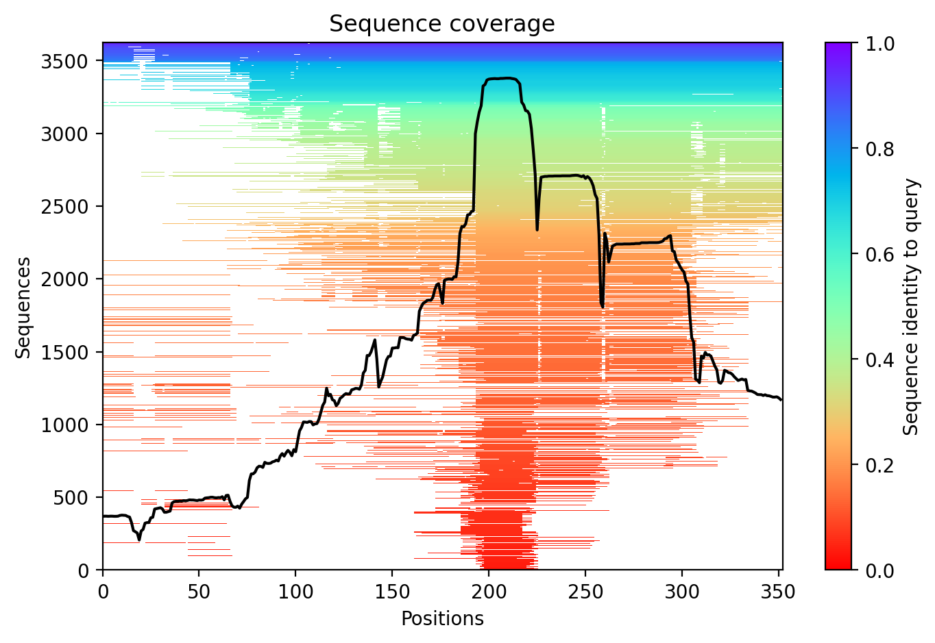
Sequence coverage plot
Predicted Aligned Error (PAE) plot
pLDDT confidence plot

05/Domain Annotations

Structural Domains & Regions

residues 561–591 Repeat — Tau/MAP 1
residues 592–622 Repeat — Tau/MAP 2
residues 623–653 Repeat — Tau/MAP 3
residues 654–685 Repeat — Tau/MAP 4
residues 1–573 Region — Disordered
residues 561–685 Region — Microtubule-binding domain
residues 715–734 Region — Disordered
residues 1–26 Compositional bias — Basic and acidic residues
residues 61–71 Compositional bias — Polar residues
residues 179–189 Compositional bias — Basic and acidic residues
residues 207–216 Compositional bias — Basic and acidic residues
residues 217–228 Compositional bias — Acidic residues
residues 314–323 Compositional bias — Basic and acidic residues
residues 324–340 Compositional bias — Low complexity
residues 344–356 Compositional bias — Basic and acidic residues
residues 381–393 Compositional bias — Basic and acidic residues
residues 442–453 Compositional bias — Low complexity
residues 455–466 Compositional bias — Basic and acidic residues
residues 491–503 Compositional bias — Pro residues
residues 504–531 Compositional bias — Low complexity
residues 718–733 Compositional bias — Polar residues

Binding Partners

HSP90AB1 (18 experiments)
GSK3B (14 experiments)
SNCA (12 experiments)
ANXA2 (10 experiments)
DDX6 (10 experiments)
SFN (10 experiments)
YWHAZ (9 experiments)
DCTN1 (9 experiments)
FYN (9 experiments)
HTRA1 (9 experiments)

Gene Ontology

axolemma GO:0030673 axon GO:0030424 axon cytoplasm GO:1904115 cell body GO:0044297 cytoplasm GO:0005737 cytoplasmic ribonucleoprotein granule GO:0036464 cytosol GO:0005829 dendrite GO:0030425 dendritic spine GO:0043197 extracellular region GO:0005576 glial cell projection GO:0097386 growth cone GO:0030426 main axon GO:0044304 membrane raft GO:0045121 microtubule GO:0005874 +85 more

06/Structural Caption

TAU V337M variant shows characteristic intrinsic disorder (average pLDDT 55.0) with the microtubule-binding domain representing the primary structured element among predominantly disordered regions.

Average pLDDT of 55.0 with only 19% (68/352) high-confidence residues indicates a predominantly disordered protein. The microtubule-binding domain (residues 561-685) shows relatively higher confidence, while N-terminal and flanking regions remain highly destabilized.

The four tandem Tau/MAP repeats (residues 561-685) constitute the microtubule-binding domain and represent the most structured region. Extensive disordered regions (residues 1-573, 715-734) and low complexity segments align with the low overall confidence, consistent with Tau's intrinsically disordered nature.

The V337M mutation occurs within a low complexity region (residues 324-340) outside the microtubule-binding domain. This methionine substitution in an already disordered segment likely has minimal impact on the limited structural elements present, though it may affect aggregation propensity.

07/Peptide Therapeutics

Aggregation Analysis

Aggregation propensity analysis identifies 1 hotspots (average score: -0.19) using Pawar+KyteDoolittle+charge algorithm.

Residues 542–546 (0.60)

08/Known Inhibitors

Known Binders from ChEMBL

CHEMBL2036430 Ki: 0.48 nM (pChEMBL 9.32)

CHEMBL2036430

CHEMBL2203439 Kd: 0.7 nM (pChEMBL 9.15)

CHEMBL2203439

CHEMBL3286988 IC50: 1.0 nM (pChEMBL 9.0)

CHEMBL3286988

CHEMBL2203332 IC50: 1.41 nM (pChEMBL 8.85)

CHEMBL2203332

CHEMBL2181533 IC50: 2.0 nM (pChEMBL 8.7)

CHEMBL2181533

CHEMBL2181532 IC50: 2.0 nM (pChEMBL 8.7)

CHEMBL2181532

CHEMBL3286982 IC50: 2.0 nM (pChEMBL 8.7)

CHEMBL3286982

CHEMBL3286983 IC50: 2.0 nM (pChEMBL 8.7)

CHEMBL3286983

CHEMBL3286984 IC50: 2.0 nM (pChEMBL 8.7)

CHEMBL3286984

CHEMBL480 Ki: 2.5 nM (pChEMBL 8.6)

LANSOPRAZOLE

09/Candidate Peptides

De Novo Peptide Design Pipeline

Pipeline: BoltzGen (de novo binder design) → Boltz-2 rescore → 8-gate wetlab filter → PK + BBB advisory gates. Target site selected from UniProt curated annotations, P2Rank pocket prediction, and aggregation propensity (in that priority order). Advisory gates annotate each candidate with estimated serum half-life, renal/immunogenicity risk, and (for CNS targets) a recommended blood-brain-barrier shuttle conjugation — without silently dropping designs.

Loading candidate statistics...

Sequences are withheld pending IP review. Full candidate data (sequences, scores, CIF files) is available to authorized reviewers via the /api/private/candidates/{fold_id} endpoint with X-Private-Key.

Legacy candidates (charge-complementary)

Target Region

Residues 542–546 (0.60 aggregation score)

Candidate ID

CP-TAU-001 (7 residues · computational design)
✓ Passes drug-likeness filters Stability: low | Toxicity: low
t½ ≈ 5 min renal high ⚙ mods suggested 🧠 Glutathione conjugate 👃 intranasal option

10/Agent Findings

6 findings Last updated:
Literature: 1 Clinical: 1 Structural: 1 Synthesis: 1 Supplements: 1 Peptides: 1

Literature Agent (1)

Literature Agent

None of the provided papers directly address the TAU V337M variant specifically. While several papers discuss tau pathology mechanisms, genetic risk factors, and biomarkers relevant to Alzheimer's disease pathogenesis, they focus on other variants (P301L, PSEN1 mutations, APOE variants, PRNP variants) or general tau biology. These papers provide contextual understanding of tau-mediated neurodegeneration but do not offer specific insights into the V337M variant's pathogenic mechanisms or clinical associations.

Clinical Agent (1)

Clinical Agent

No summary available

Structural Agent (1)

Structural Agent

AlphaFold structure update: Baseline check: 9 structure(s) found

Supplements Agent (1)

Supplements Agent

The therapeutic landscape for TAU V337M in AD includes primarily nutritional supplements (melatonin, tricaprilin) being tested in Phase 2-3 trials for their effects on tau biomarkers and cognition, alongside emerging peptide-based approaches. High-throughput screening and peptide engineering studies have identified cyclic peptides and small molecules that target tau interactions (tau-LRP1, CAPON) and tau seeding (PHF6 peptide), though these remain in preclinical stages. The BCG vaccine trial represents an immunomodulatory approach targeting AD biomarkers including tau, while silkworm pupa powder trials test protein-rich nutritional interventions.

Synthesis Agent (1)

Synthesis Agent

Synthesis of 5 findings (clinical, literature, peptides, structural, supplements): The TAU V337M variant associated with Alzheimer's disease presents a multifaceted therapeutic landsc...

Peptide Agent (1)

Peptide Agent

TAU V337M: 10 known binders (top: 0.5 nM); 1 candidate peptides designed