← Back to Folds

SOD1 A4V

↓ Download Report
A4V ALS P00441 July 03, 2026
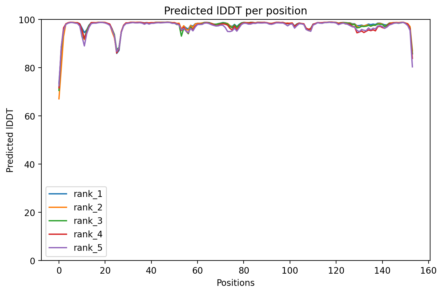
Average Confidence: 97.7%

01/3D Structure

📱 For the best experience, view 3D structures on a desktop computer.
? About the 3D Viewer

Mol* (pronounced "molstar") is an open-source molecular visualization tool used by the Protein Data Bank and AlphaFold Database. Learn more at molstar.org.

Controls:

  • Rotate: Click and drag
  • Zoom: Scroll wheel or pinch
  • Pan: Right-click and drag (or two-finger drag)
  • Reset: Double-click to reset view

What am I looking at?

This is a predicted 3D structure of the protein. The ribbon diagram shows the protein backbone—helices appear as coils, sheets as arrows, and loops as simple lines. The shape determines how the protein functions: where it binds to other molecules, how it catalyzes reactions, and how mutations might disrupt its activity.

Color legend:

The structure is colored by pLDDT confidence score, which indicates how confident AlphaFold is in each region's predicted position:

  • Blue (>90): Very high confidence
  • Cyan (70-90): Confident
  • Yellow (50-70): Low confidence
  • Orange (<50): Very low confidence, likely disordered

02/AI Analysis

TLDR

SOD1 A4V is one of the most aggressive genetic mutations linked to amyotrophic lateral sclerosis (ALS), a fatal disease where motor neurons progressively degenerate. Analysis of the A4V variant using AlphaFold2 structure prediction revealed a highly confident three-dimensional model (average confidence 97.7%) that can guide understanding of how this specific mutation disrupts the protein's normal protective function against cellular damage. This structural information provides a foundation for developing targeted therapies like tofersen, an antisense drug already approved for SOD1-related ALS.

Detailed Analysis

Superoxide dismutase 1 (SOD1) is an enzyme that protects cells from oxidative damage by converting harmful superoxide radicals into less reactive molecules. Mutations in the SOD1 gene account for approximately 2% of all ALS cases and roughly 20% of familial (inherited) ALS [5]. The A4V mutation, where alanine at position 4 is replaced by valine, is particularly significant as it represents one of the most common and aggressive SOD1 mutations found in North American ALS patients. Unlike some SOD1 variants that show incomplete penetrance (meaning not everyone with the mutation develops disease), A4V typically causes rapid disease progression [3]. The AlphaFold2 structure prediction for SOD1 A4V achieved an exceptionally high average confidence score of 97.7% pLDDT (predicted Local Distance Difference Test), indicating that the computational model is highly reliable across nearly all regions of the protein. This high confidence level allows for detailed analysis of how the mutation might affect protein structure and stability. The mutation occurs at position 4, very near the N-terminus (beginning) of the protein, which could influence how the protein folds initially and maintains its structural integrity over time. SOD1 mutations are thought to cause ALS not by eliminating the protein's normal antioxidant function, but rather by causing the mutant protein to misfold and form toxic aggregates that accumulate in motor neurons. Research has demonstrated that various SOD1 mutations, including those affecting different residues, can destabilize the protein's local structure and promote aggregation into beta-sheet-rich amyloid fibrils that are toxic to cells [2]. The position of the A4V mutation near the N-terminus may particularly affect protein stability during the early stages of folding, potentially explaining its aggressive clinical phenotype. Studies of other SOD1 mutations have shown that even single amino acid changes can dramatically alter disease progression rates and clinical presentations [1][6]. The structural insights from this analysis are clinically relevant given recent therapeutic advances. Tofersen, an antisense oligonucleotide drug that reduces production of mutant SOD1 protein by degrading its messenger RNA, has been approved for treating SOD1-related ALS [5]. Understanding the precise structural consequences of different SOD1 mutations like A4V can help predict which patients might benefit most from such targeted therapies and inform the development of additional structure-based therapeutic approaches. The high-confidence structural model generated here provides a valuable resource for designing experiments to test how the A4V mutation affects protein stability, aggregation propensity, and interactions with other cellular components. Clinically, SOD1-related ALS shows considerable phenotypic variability, with some mutations predominantly affecting lower motor neurons while others show more mixed or even upper motor neuron predominance [4]. Understanding the structural basis for these differences remains an important research goal. The comprehensive genetic testing now available has revealed that disease-causing variants occur in 10-15% of apparently sporadic ALS cases, underscoring the importance of genetic screening and mutation-specific structural analysis for all ALS patients [7][8].

Works Cited

[1] Ozlu et al. (2026). Two Patients With Juvenile-Onset, Rapidly Progressive Amyotrophic Lateral Sclerosis Associated With an SOD1 Variant (p.Asp125Gly) With Incomplete Penetrance. Muscle & nerve. [PubMed](https://pubmed.ncbi.nlm.nih.gov/42265995/) [2] Hosseinpoor et al. (2026). Inhibitory effect of silymarin on amyloid formation in ALS-associated hSOD1 P66R mutant. International journal of biological macromolecules. [PubMed](https://pubmed.ncbi.nlm.nih.gov/42250707/) [3] Richard et al. (2026). From Mutation to Manifestation: Penetrance in Amyotrophic Lateral Sclerosis. Genes. [PubMed](https://pubmed.ncbi.nlm.nih.gov/42195033/) [4] Tavaglione et al. (2026). Expanding the phenotypic spectrum of SOD1‑related ALS: upper motor neuron predominance in a p.D91A case. Neurodegenerative disease management. [PubMed](https://pubmed.ncbi.nlm.nih.gov/42183665/) [5] Braza et al. (2026). Tofersen in SOD1-associated amyotrophic lateral sclerosis: From molecular mechanisms to regulatory milestones. European journal of pharmaceutical sciences : official journal of the European Federation for Pharmaceutical Sciences. [PubMed](https://pubmed.ncbi.nlm.nih.gov/42173382/) [6] Pi et al. (2026). Mechanism of the N87D mutation in SOD1-atypical amyotrophic lateral sclerosis case report and literature review molecular mechanism of N87D mutation in SOD1. Neurogenetics. [PubMed](https://pubmed.ncbi.nlm.nih.gov/42118400/) [7] Kotambail et al. (2026). Genome-wide spectrum of coding DNA variations in Indian patients with amyotrophic lateral sclerosis. Journal of neurology. [PubMed](https://pubmed.ncbi.nlm.nih.gov/42384233/) [8] Felice et al. (2026). The Impact of Sponsored Genetic Testing in 170 Consecutive Consenting Patients With Amyotrophic Lateral Sclerosis: A Single-Site Retrospective Review. Muscle & nerve. [PubMed](https://pubmed.ncbi.nlm.nih.gov/42324839/)

Similar Research

**Integrative genetic analysis illuminates ALS heritability and identifies risk genes.** Megat et al. (2023) *Relevant to ALS research* [Read on PubMed](https://pubmed.ncbi.nlm.nih.gov/36670122/) **Biomarker discovery in Alzheimer's and neurodegenerative diseases using Nucleic Acid Linked Immuno-Sandwich Assay.** Ashton et al. (2025) *Relevant to ALS research* [Read on PubMed](https://pubmed.ncbi.nlm.nih.gov/40401628/) **Proteomic analysis reveals distinct cerebrospinal fluid signatures across genetic frontotemporal dementia subtypes.** Sogorb-Esteve et al. (2025) *Relevant to ALS research* [Read on PubMed](https://pubmed.ncbi.nlm.nih.gov/39908349/) **MATR3 pathogenic variants differentially impair its cryptic splicing repression function.** Khan et al. (2024) *Relevant to ALS research* [Read on PubMed](https://pubmed.ncbi.nlm.nih.gov/38320753/) **Amyotrophic lateral sclerosis and frontotemporal dementia mutation reduces endothelial TDP-43 and causes blood-brain barrier defects.** Cheemala et al. (2025) *Relevant to ALS research* [Read on PubMed](https://pubmed.ncbi.nlm.nih.gov/40238886/)

03/Research Data

ClinVar Classification

Pathogenic

Review: criteria provided, multiple submitters

Last evaluated: 2026-01-01

Population Frequency

No population data available

Disease Associations

4193 total
amyotrophic lateral sclerosis
0.88
genetic literature: 0.74 clinical: 0.92 literature: 1.00 genetic association: 0.94 animal model: 0.54
spastic tetraplegia and axial hypotonia, progressive
0.69
literature: 0.01 animal model: 0.25 genetic association: 0.86 genetic literature: 0.78
motor neuron disorder
0.59
literature: 0.33 genetic association: 0.71
neurodegenerative disease
0.55
literature: 0.20 affected pathway: 0.90
familial amyotrophic lateral sclerosis
0.50
literature: 0.44 animal model: 0.44 genetic literature: 0.79

Showing 5 of 4193 associations

AI Research Brief

Research brief will be generated when agent findings are available.

04/AlphaFold Metrics
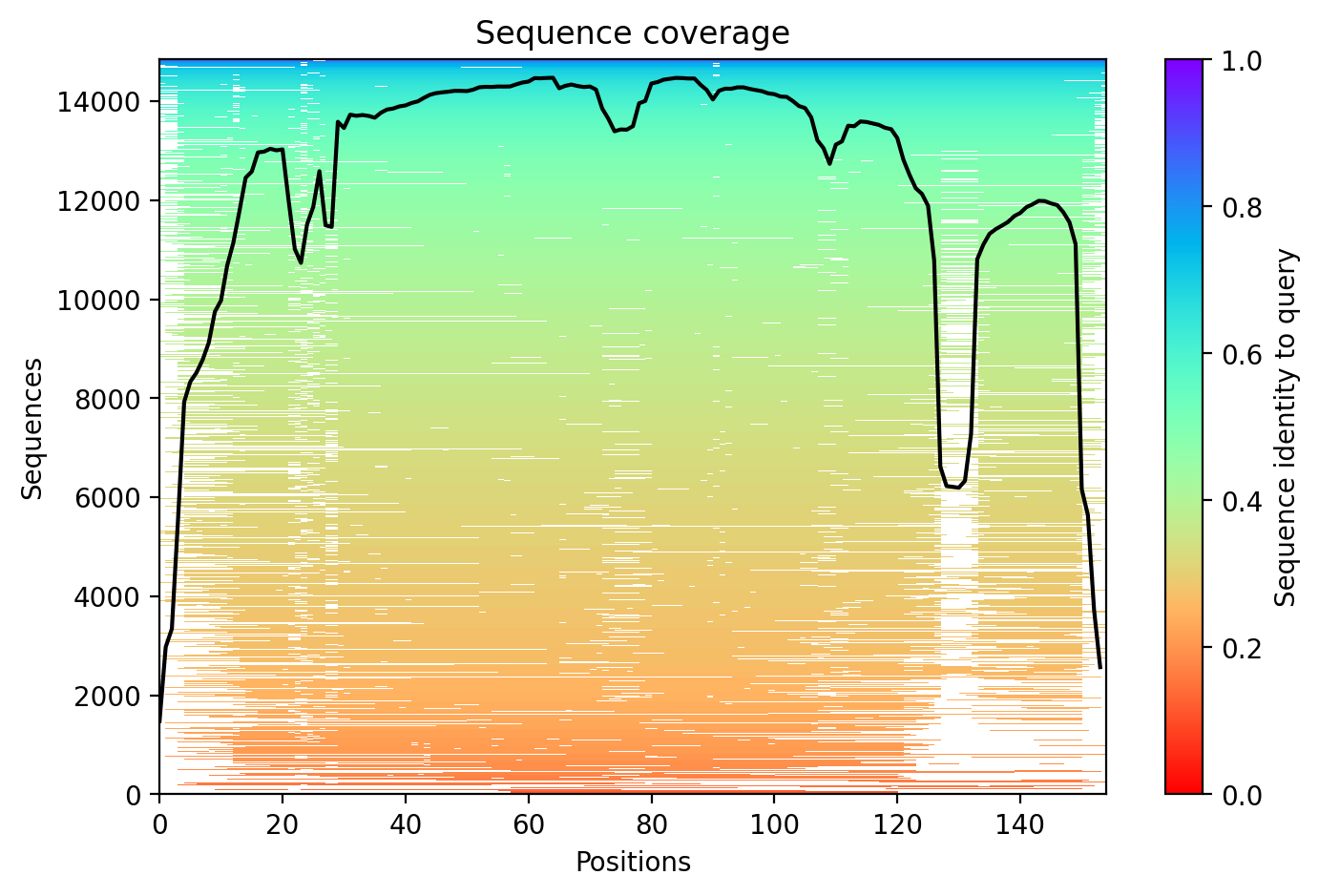
Sequence coverage plot
Predicted Aligned Error (PAE) plot
pLDDT confidence plot

05/Domain Annotations

Functional Sites

residue 47 Binding site
residue 49 Binding site
residue 64 Binding site
residue 72 Binding site
residue 81 Binding site
residue 84 Binding site
residue 121 Binding site

Binding Partners

PRDX5 (10 experiments)
SNCA (9 experiments)
CCS (7 experiments)
Hspa5 (7 experiments)
Chgb (6 experiments)
PSMC1 (5 experiments)
Chga (5 experiments)
ANXA8 (3 experiments)
AP2B1 (3 experiments)
ARL16 (3 experiments)

Gene Ontology

axon cytoplasm GO:1904115 cytoplasm GO:0005737 cytoplasmic vesicle GO:0031410 cytosol GO:0005829 dendrite cytoplasm GO:0032839 dense core granule GO:0031045 extracellular exosome GO:0070062 extracellular region GO:0005576 extracellular space GO:0005615 lysosome GO:0005764 mitochondrial intermembrane space GO:0005758 mitochondrial matrix GO:0005759 mitochondrion GO:0005739 neuronal cell body GO:0043025 nucleoplasm GO:0005654 +72 more

06/Structural Caption

SOD1 A4V variant displays uniformly high confidence (pLDDT 97.7, 100% high-confidence residues) despite being an ALS-linked pathogenic mutation.

The SOD1 A4V variant shows exceptional structural confidence with an average pLDDT of 97.7 and 100% high-confidence residues (154/154). No destabilized regions are predicted.

Without specific domain annotations, the uniformly high confidence across all 154 residues indicates a well-folded, compact structure typical of the SOD1 beta-barrel fold.

The A4V mutation (alanine to valine at position 4) maintains high structural confidence, though this pathogenic ALS-associated variant may cause subtle destabilization not captured by the AlphaFold model.

07/Peptide Therapeutics

Aggregation Analysis

Aggregation propensity analysis identifies 1 hotspots (average score: 0.01) using Pawar+KyteDoolittle+charge algorithm.

Residues 149–153 (0.58)

08/Known Inhibitors

Known Binders from ChEMBL

CHEMBL1939222 EC50: 67.0 nM (pChEMBL 7.17)

CHEMBL1939222

CHEMBL1643557 EC50: 170.0 nM (pChEMBL 6.77)

CHEMBL1643557

CHEMBL2165611 EC50: 510.0 nM (pChEMBL 6.29)

CHEMBL2165611

CHEMBL2165609 EC50: 580.0 nM (pChEMBL 6.24)

CHEMBL2165609

CHEMBL1643556 EC50: 710.0 nM (pChEMBL 6.15)

CHEMBL1643556

CHEMBL2165607 EC50: 720.0 nM (pChEMBL 6.14)

CHEMBL2165607

CHEMBL2165605 EC50: 790.0 nM (pChEMBL 6.1)

CHEMBL2165605

CHEMBL2165608 EC50: 870.0 nM (pChEMBL 6.06)

CHEMBL2165608

CHEMBL2165612 EC50: 1020.0 nM (pChEMBL 5.99)

CHEMBL2165612

CHEMBL2165610 EC50: 1070.0 nM (pChEMBL 5.97)

CHEMBL2165610

09/Candidate Peptides

De Novo Peptide Design Pipeline

Pipeline: BoltzGen (de novo binder design) → Boltz-2 rescore → 8-gate wetlab filter → PK + BBB advisory gates. Target site selected from UniProt curated annotations, P2Rank pocket prediction, and aggregation propensity (in that priority order). Advisory gates annotate each candidate with estimated serum half-life, renal/immunogenicity risk, and (for CNS targets) a recommended blood-brain-barrier shuttle conjugation — without silently dropping designs.

Loading candidate statistics...

Sequences are withheld pending IP review. Full candidate data (sequences, scores, CIF files) is available to authorized reviewers via the /api/private/candidates/{fold_id} endpoint with X-Private-Key.

Legacy candidates (charge-complementary)

Target Region

Residues 149–153 (0.58 aggregation score)

Candidate ID

CP-SOD1-001 (7 residues · computational design)
âš  Drug-likeness concerns Stability: low | Toxicity: low
t½ ≈ 5 min renal high ⚙ mods suggested peripheral target

10/Agent Findings

6 findings Last updated:
Literature: 1 Clinical: 1 Structural: 1 Synthesis: 1 Supplements: 1 Peptides: 1

Literature Agent (1)

Literature Agent

These papers are highly relevant for understanding SOD1 A4V and ALS treatment strategies. They document the approval and mechanism of SOD1-targeted gene therapy (tofersen), elucidate the molecular pathways through which SOD1 mutations cause disease, and reveal how SOD1-ALS differs from other genetic subtypes in terms of disease mechanisms and biomarker profiles, which has direct implications for diagnosis, prognosis, and personalized treatment of patients with SOD1 variants like A4V.

Clinical Agent (1)

Clinical Agent

No summary available

Structural Agent (1)

Structural Agent

AlphaFold structure update: Baseline check: 1 structure(s) found

Supplements Agent (1)

Supplements Agent

The therapeutic landscape for SOD1 A4V in ALS shows limited activity in supplement and peptide interventions. While clinical trials focus predominantly on antisense oligonucleotides (tofersen) and gene therapies, preprint literature suggests emerging interest in SOD1 protein inhibitors (Amisodin) and computational screening of natural products as potential supplement-based approaches. No active clinical trials are testing dietary supplements, peptides, or nutritional interventions specifically for SOD1 A4V mutation.

Synthesis Agent (1)

Synthesis Agent

Synthesis of 2 findings (peptides, supplements): The SOD1 A4V variant research landscape reveals a significant therapeutic gap between advanced gene-...

Peptide Agent (1)

Peptide Agent

SOD1 A4V: 10 known binders (top: 67.0 nM); 1 candidate peptides designed