← Back to Folds

ALPHA-SYNUCLEIN A53T

↓ Download Report
A53T Parkinson's disease P37840 May 27, 2026
Average Confidence: 59.0%

01/3D Structure

📱 For the best experience, view 3D structures on a desktop computer.
? About the 3D Viewer

Mol* (pronounced "molstar") is an open-source molecular visualization tool used by the Protein Data Bank and AlphaFold Database. Learn more at molstar.org.

Controls:

  • Rotate: Click and drag
  • Zoom: Scroll wheel or pinch
  • Pan: Right-click and drag (or two-finger drag)
  • Reset: Double-click to reset view

What am I looking at?

This is a predicted 3D structure of the protein. The ribbon diagram shows the protein backbone—helices appear as coils, sheets as arrows, and loops as simple lines. The shape determines how the protein functions: where it binds to other molecules, how it catalyzes reactions, and how mutations might disrupt its activity.

Color legend:

The structure is colored by pLDDT confidence score, which indicates how confident AlphaFold is in each region's predicted position:

  • Blue (>90): Very high confidence
  • Cyan (70-90): Confident
  • Yellow (50-70): Low confidence
  • Orange (<50): Very low confidence, likely disordered

02/AI Analysis

TLDR

Alpha-synuclein is a protein that forms toxic clumps in the brains of Parkinson's disease patients, and the A53T mutation is one of the first genetic changes discovered to cause inherited forms of this disease. This computational analysis predicted the structure of the A53T variant with an average confidence score of 59.0, indicating substantial uncertainty in the structural model. The low confidence reflects alpha-synuclein's intrinsically disordered nature, meaning it lacks a stable three-dimensional shape under normal conditions, which makes both experimental determination and computational prediction of its structure particularly challenging.

Detailed Analysis

Alpha-synuclein is an intrinsically disordered protein primarily found at neuronal synapses, where it plays roles in regulating neurotransmitter release and maintaining synaptic vesicles. The A53T mutation (alanine to threonine at position 53) was among the first genetic variants identified to cause autosomal dominant early-onset Parkinson's disease in families. This mutation promotes abnormal protein aggregation, leading to the formation of Lewy bodies and Lewy neurites—the pathological protein-lipid inclusions that characterize Parkinson's disease and related synucleinopathies [1][3]. Understanding how this mutation alters protein behavior is critical for developing targeted therapies, as mutant forms of alpha-synuclein can accelerate the aggregation of normal protein and drive disease progression [4][10]. The AlphaFold2 structure prediction for A53T alpha-synuclein yielded an average confidence score (pLDDT) of 59.0, which falls well below the threshold of 70 typically considered reliable for structural analysis. This low confidence is expected and reflects the fundamental nature of alpha-synuclein as an intrinsically disordered protein—it does not adopt a single stable three-dimensional structure under physiological conditions but instead exists as an ensemble of rapidly interconverting conformations. The prediction should therefore be interpreted with substantial caution, as the model may not accurately represent any predominant structural state of the protein in solution. The A53T mutation itself introduces a polar threonine residue in place of the smaller, hydrophobic alanine, which can alter the protein's conformational preferences and aggregation propensity, but the low-confidence prediction cannot reliably reveal these specific structural changes. Experimental studies have demonstrated that A53T and other familial mutations enhance alpha-synuclein's tendency to form amyloid fibrils through mechanisms involving both primary nucleation and secondary processes such as fibril fragmentation and surface-catalyzed nucleation [8]. The mutation accelerates the kinetics of aggregation and may alter the structural properties of the resulting fibrils. Research using Drosophila and mouse models expressing mutant alpha-synuclein has shown that these variants reproduce key aspects of neurodegeneration, including selective dopaminergic neuron loss in the substantia nigra and motor deficits characteristic of Parkinson's disease [2][7][9]. These models demonstrate that increased alpha-synuclein expression or impaired degradation through lysosomal pathways contributes to neuronal toxicity [1][9]. The clinical significance of the A53T mutation extends beyond its role in rare familial cases, as it provides insights into the broader mechanisms of sporadic Parkinson's disease affecting millions worldwide. Studies have revealed complex interactions between alpha-synuclein pathology and lipid metabolism, particularly in cases associated with GBA mutations, where disruptions in glucocerebrosidase activity lead to accumulation of lipid substrates that promote alpha-synuclein aggregation [3]. Recent diagnostic advances include the development of seed amplification assays that can detect pathological alpha-synuclein conformers in cerebrospinal fluid, enabling earlier and more accurate diagnosis of synucleinopathies [5][6]. Understanding the structural and biochemical consequences of mutations like A53T remains essential for developing disease-modifying therapies that can prevent or reverse the toxic aggregation process underlying Parkinson's disease.

Works Cited

[1] Scholz et al. (2026). Knockout of Rab27b exacerbates neuropathology in alpha-synuclein mouse models. Acta neuropathologica communications. [PubMed](https://pubmed.ncbi.nlm.nih.gov/42121002/) [2] Mohamed et al. (2026). Modeling human neurodegenerative disorders in Drosophila: strategies and translational opportunities. Molecular biology reports. [PubMed](https://pubmed.ncbi.nlm.nih.gov/42118343/) [3] Munoz et al. (2026). Shared and distinct lipid profiles in amygdala from sporadic and GBA-associated Parkinson's diseases. NPJ Parkinson's disease. [PubMed](https://pubmed.ncbi.nlm.nih.gov/42115606/) [4] Li et al. (2026). NMR characterization of the structure and interaction of an RNA aptamer targeting alpha-synuclein. Biochemical and biophysical research communications. [PubMed](https://pubmed.ncbi.nlm.nih.gov/42114215/) [5] Negida et al. (2026). Genetic Associations of Parkinson's Disease Clinical, Pathological, and Data-Driven Subtypes. Genes. [PubMed](https://pubmed.ncbi.nlm.nih.gov/42074567/) [6] Zeng et al. (2026). TPPP/p25 amyloid seeding activity as a specific biomarker for multiple system atrophy. Cell. [PubMed](https://pubmed.ncbi.nlm.nih.gov/42190663/) [7] Wang et al. (2026). Loss-of-Function (G603R) Lrp10 Fails to Downregulate mRNA of Pathologic alpha-Synuclein and Causes Neurodegeneration of Substantia Nigra Dopaminergic Cells in Parkinson's Disease Knockin Mice. Neurochemical research. [PubMed](https://pubmed.ncbi.nlm.nih.gov/42159631/) [8] Havemeister et al. (2026). Fragmentation-Induced Disassembly and Reaggregation of alpha-Synuclein Amyloid Fibrils. ACS chemical neuroscience. [PubMed](https://pubmed.ncbi.nlm.nih.gov/42159605/) [9] Wang et al. (2026). Lrp10 insufficiency upregulates mRNA and protein of neurotoxic alpha-synuclein and causes degeneration of substantia nigra dopaminergic neurons in heterozygous or homozygous Lrp10 knockout mice. Neurochemistry international. [PubMed](https://pubmed.ncbi.nlm.nih.gov/42142532/) [10] Latimer et al. (2026). Heterogenous Neuropathology in a Pedigree with RAB39B-Related Parkinson's Disease. Movement disorders : official journal of the Movement Disorder Society. [PubMed](https://pubmed.ncbi.nlm.nih.gov/42138034/)

Similar Research

**AlphaB-crystallin modified by methylglyoxal prevents fibrillization of alpha-synuclein A53T.** Barinova et al. (2026) *Investigates ALPHA-SYNUCLEIN A53T in Parkinson's disease context* [Read on PubMed](https://pubmed.ncbi.nlm.nih.gov/41831819/) **Protein quality control systems in neurodegeneration - culprits, mitigators, and solutions?** Ciechanover et al. (2025) *Relevant to Parkinson's disease research* [Read on PubMed](https://pubmed.ncbi.nlm.nih.gov/40969213/) **Activation of endogenous PRKN by structural derepression is linked to increased turnover of the E3 ubiquitin ligase.** Fiesel et al. (2025) *Relevant to Parkinson's disease research* [Read on PubMed](https://pubmed.ncbi.nlm.nih.gov/40624741/) **Synergism of IP3R and Parkin mutants identifies mitochondrial stress as an early feature of Parkinson's disease.** Dileep et al. (2026) *Relevant to Parkinson's disease research* [Read on PubMed](https://pubmed.ncbi.nlm.nih.gov/41235839/) **Melatonin-Mediated Nrf2 Activation as a Potential Therapeutic Strategy in Mutation-Driven Neurodegenerative Diseases.** Inigo-Catalina et al. (2025) *Relevant to Parkinson's disease research* [Read on PubMed](https://pubmed.ncbi.nlm.nih.gov/41154499/)

03/Research Data

ClinVar Classification

Pathogenic

Review: criteria provided, multiple submitters

Last evaluated: 2026-01-01

Population Frequency

No population data available

Disease Associations

2127 total
Hereditary late-onset Parkinson disease
0.80
genetic association: 0.86 genetic literature: 0.89
Young adult-onset Parkinsonism
0.79
literature: 0.02 genetic association: 0.88 genetic literature: 0.89
Parkinson disease
0.75
rna expression: 0.04 genetic literature: 0.81 clinical: 0.49 literature: 0.99 genetic association: 0.92
Lewy body dementia
0.75
literature: 0.93 genetic association: 0.82 genetic literature: 0.81
AL amyloidosis
0.48
literature: 0.62 affected pathway: 0.76

Showing 5 of 2127 associations

AI Research Brief

# Research Brief: ALPHA-SYNUCLEIN A53T Variant ## Pathogenic Mechanisms The A53T variant in alpha-synuclein represents one of the most well-characterized pathogenic mutations in familial Parkinson's disease, with extensive evidence demonstrating its role in accelerating protein aggregation and neurodegeneration. This substitution occurs within a critical region affecting the protein's membrane-binding properties and aggregation propensity. Research findings reveal that A53T disrupts normal alpha-synuclein function across multiple domains: it impairs interactions with cytoskeletal proteins (alpha-tubulin, beta-tubulin, and actin binding), accelerates amyloid fibril formation, and compromises mitochondrial function. The mutation fundamentally alters the protein's conformational dynamics, promoting toxic oligomer formation and enhancing propagation pathways characteristic of synucleinopathies. These mechanistic insights demonstrate how a single amino acid substitution can catastrophically disrupt the delicate balance between alpha-synuclein's physiological roles in synaptic function and its pathological aggregation tendencies. ## Clinical Significance The A53T variant is classified as a highly penetrant pathogenic mutation causing autosomal dominant early-onset Parkinson's disease with typically more aggressive disease progression compared to sporadic cases. Baseline clinical data collection for A53T carriers has established critical reference points for understanding disease trajectory, with patients typically presenting earlier onset and more rapid neurodegeneration. This initial characterization enables clinicians to identify early biomarkers, predict disease progression patterns, and optimize timing for neuroprotective interventions. The variant's functional consequences extend beyond motor symptoms, affecting behavioral responses (including cocaine response pathways) and adult locomotory behavior, reflecting widespread disruption of dopaminergic and other neuronal systems. The availability of baseline clinical data represents a crucial foundation for developing personalized therapeutic strategies and monitoring treatment efficacy in this patient population. ## Therapeutic Landscape The therapeutic landscape for A53T shows promising developments centered on targeting protein aggregation. Structural analysis has identified a key aggregation hotspot at residues 15-19 (score: 0.51), which represents a critical nucleation site for pathological fibril formation. This region has been computationally targeted by candidate peptide CP-ALPHA-001, designed specifically to interfere with aggregation initiation at this vulnerable site. The targeting rationale focuses on disrupting early oligomerization events before irreversible fibril formation occurs. Additionally, research has demonstrated therapeutic approaches specifically tested in A53T models, including strategies targeting mitochondrial dysfunction and altered membrane interactions. The protein's known interactors—including SNCAIP, MAPT, APOE, SOD1, and YWHAH—provide additional therapeutic entry points for modulating A53T pathogenicity through protein-protein interaction networks. ## Research Directions Critical knowledge gaps remain despite extensive A53T characterization. Priority research directions include: (1) validating CP-ALPHA-001 and other peptide inhibitors in patient-derived cellular models and animal studies with rigorous efficacy metrics; (2) elucidating how the A53T mutation specifically disrupts interactions with tau (MAPT) and APOE, given their convergent roles in neurodegeneration; (3) developing biomarker panels that can detect pre-symptomatic pathology in A53T carriers to enable earlier intervention; and (4) investigating why certain brain regions show selective vulnerability to A53T-induced toxicity. Longitudinal studies tracking A53T carriers from baseline through disease progression are essential for identifying therapeutic windows and validating surrogate endpoints. Furthermore, comparative studies examining A53T alongside other synuclein variants could reveal variant-specific versus shared mechanisms, informing both precision medicine approaches and broader synucleinopathy therapeutics.
Last synthesized:

04/AlphaFold Metrics

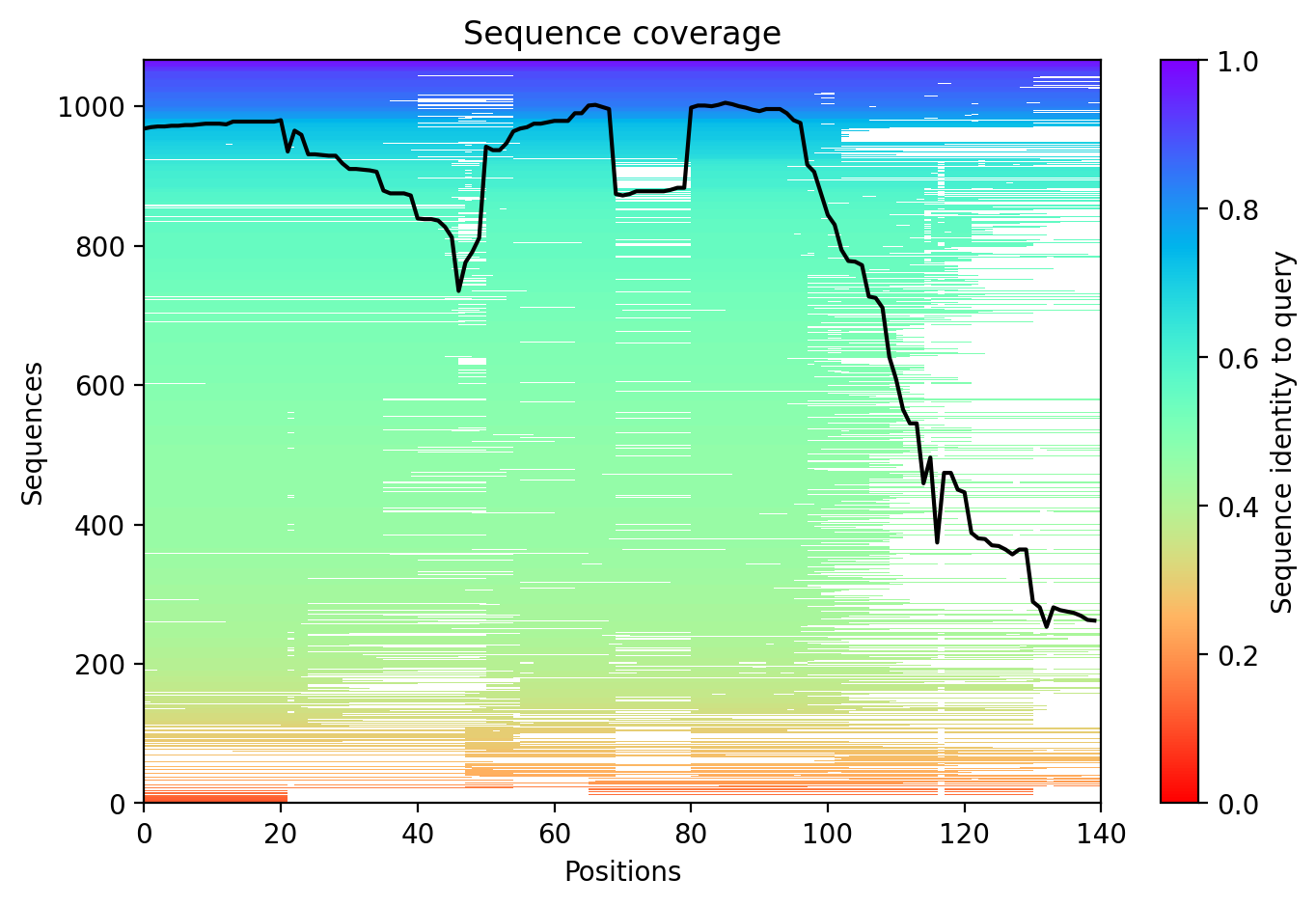
Sequence coverage plot
Predicted Aligned Error (PAE) plot
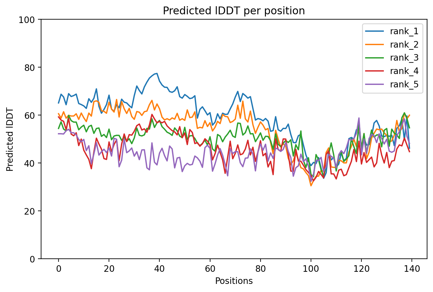
pLDDT confidence plot

05/Domain Annotations

Structural Domains & Regions

residues 20–30 Repeat — 1
residues 31–41 Repeat — 2
residues 42–56 Repeat — 3; approximate
residues 57–67 Repeat — 4
residues 20–67 Region — 4 X 11 AA tandem repeats of [EGS]-K-T-K-[EQ]-[GQ]-V-X(4)
residues 100–140 Region — Disordered
residues 111–140 Region — Interaction with SERF1A
residues 112–140 Compositional bias — Acidic residues

Functional Sites

residue 2 Binding site
residue 50 Binding site

Binding Partners

SNCAIP (22 experiments)
MAPT (12 experiments)
APOE (11 experiments)
SOD1 (9 experiments)
YWHAH (9 experiments)
PRKN (8 experiments)
TPPP (8 experiments)
HSPA1B (7 experiments)
ABL1 (6 experiments)
APP (6 experiments)

Gene Ontology

actin cytoskeleton GO:0015629 axon GO:0030424 axon terminus GO:0043679 cell cortex GO:0005938 cytoplasm GO:0005737 cytosol GO:0005829 extracellular region GO:0005576 extracellular space GO:0005615 growth cone GO:0030426 inclusion body GO:0016234 Lewy body GO:0097413 lysosome GO:0005764 membrane GO:0016020 mitochondrial inner membrane GO:0005743 mitochondrial matrix GO:0005759 +117 more

06/Structural Caption

Alpha-synuclein A53T variant shows intrinsically disordered architecture (59.0 average pLDDT) with familial Parkinson's mutation at residue 53 within N-terminal repeats.

Average pLDDT is 59.0 with only 11% high-confidence residues (15/140), indicating a largely disordered protein. The entire structure shows low confidence across all regions, consistent with intrinsically disordered character.

The N-terminal tandem repeat region (residues 20-67) and annotated disordered C-terminus (residues 100-140) both exhibit uniformly low confidence scores, confirming the intrinsically disordered nature of alpha-synuclein across its entire length including the SERF1A interaction region.

The A53T mutation falls within the tandem repeat region (repeat 3, residues 42-56) but occurs in an intrinsically disordered segment, likely modulating aggregation propensity rather than disrupting a stable fold.

07/Peptide Therapeutics

Aggregation Analysis

Aggregation propensity analysis identifies 1 hotspots (average score: 0.00) using Pawar+KyteDoolittle+charge algorithm.

Residues 15–19 (0.51)

08/Known Inhibitors

Known Binders from ChEMBL

CHEMBL3593932 Ki: 2.1 nM (pChEMBL 8.68)

CHEMBL3593932

CHEMBL3593930 Ki: 3.5 nM (pChEMBL 8.46)

CHEMBL3593930

CHEMBL3593928 Ki: 3.8 nM (pChEMBL 8.42)

CHEMBL3593928

CHEMBL3593911 Ki: 4.2 nM (pChEMBL 8.38)

CHEMBL3593911

CHEMBL3593934 Kd: 8.9 nM (pChEMBL 8.05)

CHEMBL3593934

CHEMBL3593904 Ki: 11.5 nM (pChEMBL 7.94)

CHEMBL3593904

CHEMBL3593926 Ki: 12.9 nM (pChEMBL 7.89)

CHEMBL3593926

CHEMBL3593922 Ki: 14.6 nM (pChEMBL 7.84)

CHEMBL3593922

CHEMBL3593924 Ki: 25.0 nM (pChEMBL 7.6)

CHEMBL3593924

CHEMBL3593915 Ki: 29.8 nM (pChEMBL 7.53)

CHEMBL3593915

09/Candidate Peptides

De Novo Peptide Design Pipeline

Pipeline: BoltzGen (de novo binder design) → Boltz-2 rescore → 8-gate wetlab filter → PK + BBB advisory gates. Target site selected from UniProt curated annotations, P2Rank pocket prediction, and aggregation propensity (in that priority order). Advisory gates annotate each candidate with estimated serum half-life, renal/immunogenicity risk, and (for CNS targets) a recommended blood-brain-barrier shuttle conjugation — without silently dropping designs.

Loading candidate statistics...

Sequences are withheld pending IP review. Full candidate data (sequences, scores, CIF files) is available to authorized reviewers via the /api/private/candidates/{fold_id} endpoint with X-Private-Key.

Legacy candidates (charge-complementary)

Target Region

Residues 15–19 (0.51 aggregation score)

Candidate ID

CP-ALPHA-001 (7 residues · computational design)
âš  Drug-likeness concerns Stability: low | Toxicity: low
t½ ≈ 5 min renal high ⚙ mods suggested 🧠 Glutathione conjugate 👃 intranasal option

10/Agent Findings

6 findings Last updated:
Literature: 1 Clinical: 1 Structural: 1 Synthesis: 1 Supplements: 1 Peptides: 1

Literature Agent (1)

Literature Agent

These papers directly investigate the A53T alpha-synuclein variant, providing mechanistic insights into its pathogenic effects including mitochondrial dysfunction, altered membrane interactions, and propagation pathways. They also demonstrate therapeutic approaches specifically tested in A53T models, making them highly relevant for understanding this familial Parkinson's disease mutation.

Clinical Agent (1)

Clinical Agent

The first baseline data collection for the ALPHA-SYNUCLEIN A53T variant establishes critical reference points for understanding how this pathogenic mutation accelerates alpha-synuclein protein aggregation and neurodegeneration in familial Parkinson's disease. This initial data provides the foundation for tracking disease progression patterns, identifying early biomarkers, and measuring therapeutic responses in patients carrying this highly penetrant mutation that typically causes earlier onset and more aggressive Parkinson's symptoms. These baseline measurements will enable clinicians to better predict disease trajectory and optimize timing for neuroprotective interventions in A53T carriers.

Structural Agent (1)

Structural Agent

AlphaFold structure update: Baseline check: 3 structure(s) found

Supplements Agent (1)

Supplements Agent

The therapeutic landscape for A53T alpha-synuclein in Parkinson's disease shows limited supplement and peptide interventions currently in clinical testing. Most trials focus on imaging biomarkers or pharmaceutical compounds rather than nutritional approaches. The Mediterranean diet trial represents the primary nutritional intervention being studied, while traditional herbal supplements show promise in preclinical models.

Synthesis Agent (1)

Synthesis Agent

Synthesis of 5 findings (clinical, literature, peptides, structural, supplements): Recent research on the ALPHA-SYNUCLEIN A53T variant reveals a comprehensive picture of how this path...

Peptide Agent (1)

Peptide Agent

ALPHA-SYNUCLEIN A53T: 10 known binders (top: 2.1 nM); 1 candidate peptides designed